Nuclear Matrix Protein p84 (THOC1) Antibody (CAB8179)
The Nuclear Matrix Protein p84 (THOC1) Antibody (CAB8179) is a high-quality antibody developed for reliable detection and analysis of target proteins. Predicted to enable DNA binding activity and RNA binding activity. Involved in several processes, including negative regulation of DNA damage checkpoint; regulation of nucleobase-containing compound metabolic process; and viral mRNA export from host cell nucleus. Located in cytoplasm and nuclear speck. Part of THO complex part of transcription export complex. Colocalizes with chromosome, telomeric region.
This antibody is validated for use in WB, IHC-P, IF/ICC, ELISA applications and has demonstrated reactivity against Human, Mouse, Rat samples.
Product Name:
Nuclear Matrix Protein p84 (THOC1) Antibody
SKU:
CAB8179
Size:
100μL, 20μL
Reactivity:
Human, Mouse, Rat
Conjugate:
Unconjugated
Immunogen:
Recombinant protein (or fragment).This information is considered to be commercially sensitive.
Tested Applications:
WBIHC-PIF/ICCELISA
Recommended Dilution:
WB
1:500 - 1:1000
IHC-P
1:50 - 1:200
IF/ICC
1:50 - 1:100
ELISA
Recommended starting concentration is 1 μg/mL. Please optimize the concentration based on your specific assay requirements.
Synonyms:
P84, HPR1, P84N5, DFNA86, Nuclear Matrix Protein p84 (THOC1)
Predicted to enable DNA binding activity and RNA binding activity. Involved in several processes, including negative regulation of DNA damage checkpoint; regulation of nucleobase-containing compound metabolic process; and viral mRNA export from host cell nucleus. Located in cytoplasm and nuclear speck. Part of THO complex part of transcription export complex. Colocalizes with chromosome, telomeric region.
Purification Method
Affinity purification
Gene ID
9984
RRID
AB_2772593
Buffer Information
Store at -20℃. Avoid freeze / thaw cycles. Buffer: PBS containing 50% glycerol, preserved with proclin300 or sodium azide, pH 7.3.
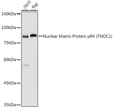
Western blot analysis of various lysates using Nuclear Matrix Protein p84 (THOC1) Rabbit pAb (CAB8179) at 1:1000 dilution. Secondary antibody: HRP-conjugated Goat anti-Rabbit IgG (H+L) (AS014) at 1:10000 dilution. Lysates/proteins: 25μg per lane. Blocking buffer: 3% nonfat dry milk in TBST. Detection: ECL Basic Kit (AbGn00020). Exposure time: 1s.
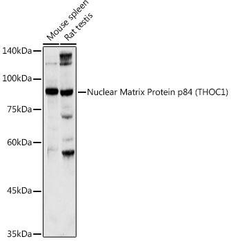
Western blot analysis of various lysates using Nuclear Matrix Protein p84 (THOC1) Rabbit pAb (CAB8179) at 1:1000 dilution. Secondary antibody: HRP-conjugated Goat anti-Rabbit IgG (H+L) (AS014) at 1:10000 dilution. Lysates/proteins: 25μg per lane. Blocking buffer: 3% nonfat dry milk in TBST. Detection: ECL Basic Kit (AbGn00020). Exposure time: 30s.
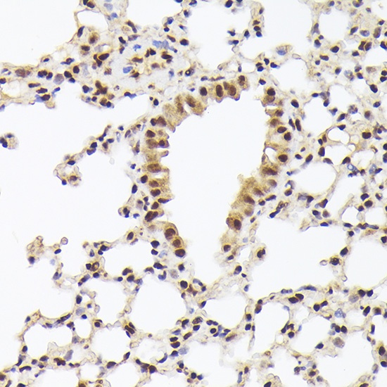
Immunohistochemistry analysis of paraffin-embedded Mouse lung using Nuclear Matrix Protein p84 (THOC1) Rabbit pAb (CAB8179) at dilution of 1:100 (40x lens). High pressure antigen retrieval performed with 0.01M Citrate buffer (pH 6.0) prior to IHC staining.
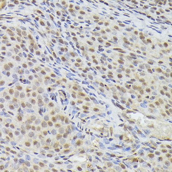
Immunohistochemistry analysis of paraffin-embedded Rat ovary using Nuclear Matrix Protein p84 (THOC1) Rabbit pAb (CAB8179) at dilution of 1:100 (40x lens). High pressure antigen retrieval performed with 0.01M Citrate buffer (pH 6.0) prior to IHC staining.