The TIM-1/KIM-1/HAVCR Polyclonal Antibody (CAB24493) is a high-quality antibody developed for reliable detection and analysis of target proteins. Enables phosphatidylserine binding activity. Acts upstream of or within positive regulation of mast cell activation; response to lipopolysaccharide; and response to wounding. Located in brush border and cell surface. Is expressed in brain; dorsal aorta; liver; lung; and reproductive system. Human ortholog(s) of this gene implicated in atopic dermatitis. Orthologous to human HAVCR1 (hepatitis A virus cellular receptor 1). RRID Gene ID 171283 Swiss Prot Synonym Tim1; KIM-1; TIM-1; Timd1; TIM-1/KIM-1/HAVCR
This antibody is validated for use in WB, ELISA applications and has demonstrated reactivity against Human samples.
Product Name:
TIM-1/KIM-1/HAVCR Polyclonal Antibody
SKU:
CAB24493
Size:
100μL, 20μL
Reactivity:
Human
Clone Number:
-
Conjugate:
Unconjugated
Immunogen:
Recombinant protein (or fragment).This information is considered to be commercially sensitive.
Tested Applications:
WBELISA
Recommended Dilution:
WB
1:2000 - 1:8000
ELISA
Recommended starting concentration is 1 μg/mL. Please optimize the concentration based on your specific assay requirements.
Synonyms:
Tim1, KIM-1, TIM-1, Timd1, TIM-1/KIM-1/HAVCR
Positive Sample:
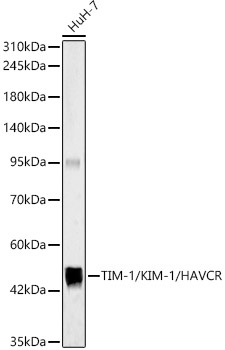
HuH-7
Cellular Localization:
-
Calculated MW:
33kDa
Observed MW:
50kDa
Enables phosphatidylserine binding activity. Acts upstream of or within positive regulation of mast cell activation; response to lipopolysaccharide; and response to wounding. Located in brush border and cell surface. Is expressed in brain; dorsal aorta; liver; lung; and reproductive system. Human ortholog(s) of this gene implicated in atopic dermatitis. Orthologous to human HAVCR1 (hepatitis A virus cellular receptor 1). RRID Gene ID 171283 Swiss Prot Synonym Tim1; KIM-1; TIM-1; Timd1; TIM-1/KIM-1/HAVCR
Purification Method:
Affinity purification
Gene ID:
171283
RRID:
-
Buffer Information:
Store at -20℃. Avoid freeze / thaw cycles. Buffer: PBS containing 50% glycerol, preserved with proclin300 or sodium azide, pH 7.3.
Western blot analysis of lysates from HuH-7 cells, using TIM-1/KIM-1/HAVCR Rabbit pAb (CAB24493) at 1:7000 dilution. Secondary antibody: HRP-conjugated Goat anti-Rabbit IgG (H+L) (AS014) at 1:10000 dilution. Lysates/proteins: 25μg per lane. Blocking buffer: 3% nonfat dry milk in TBST. Detection: ECL Basic Kit (AbGn00020). Exposure time: 180s.
 at 1:7000 dilution. Secondary antibody: HRP Goat Anti-Rabbit IgG (H+L) at 1:10000 dilution. Lysates/proteins: 25ug per lane. Blocking buffer: 3% nonfat dry milk in TBST.")
 at 1:7000 dilution. Secondary antibody: HRP Goat Anti-Rabbit IgG (H+L) at 1:10000 dilution. Lysates/proteins: 25ug per lane. Blocking buffer: 3% nonfat dry milk in TBST.")