The TNFAIP3 Antibody (CAB2127) is a high-quality antibody developed for reliable detection and analysis of target proteins. This gene was identified as a gene whose expression is rapidly induced by the tumor necrosis factor (TNF). The protein encoded by this gene is a zinc finger protein and ubiqitin-editing enzyme, and has been shown to inhibit NF-kappa B activation as well as TNF-mediated apoptosis. The encoded protein, which has both ubiquitin ligase and deubiquitinase activities, is involved in the cytokine-mediated immune and inflammatory responses. Several transcript variants encoding the same protein have been found for this gene.
This antibody is validated for use in WB, IHC-P, ELISA applications and has demonstrated reactivity against Human, Rat samples.
Product Name:
TNFAIP3 Antibody
SKU:
CAB2127
Size:
100μL, 20μL
Reactivity:
Human, Rat
Conjugate:
Unconjugated
Immunogen:
Recombinant protein (or fragment).This information is considered to be commercially sensitive.
Tested Applications:
WBIHC-PELISA
Recommended Dilution:
WB
1:100 - 1:500
IHC-P
1:50 - 1:200
ELISA
Recommended starting concentration is 1 μg/mL. Please optimize the concentration based on your specific assay requirements.
Synonyms:
A20, AISBL, AIFBL1, OTUD7C, TNFA1P2, TNFAIP3
Positive Sample:
THP-1 treated with TPA and LPS, Rat thymus
Cellular Localization:
Cytoplasm, Lysosome, Nucleus.
Calculated MW:
90 kDa
Observed MW:
90 kDa
This gene was identified as a gene whose expression is rapidly induced by the tumor necrosis factor (TNF). The protein encoded by this gene is a zinc finger protein and ubiqitin-editing enzyme, and has been shown to inhibit NF-kappa B activation as well as TNF-mediated apoptosis. The encoded protein, which has both ubiquitin ligase and deubiquitinase activities, is involved in the cytokine-mediated immune and inflammatory responses. Several transcript variants encoding the same protein have been found for this gene.
Purification Method
Affinity purification
Gene ID
7128
RRID
AB_2764146
Buffer Information
Store at -20℃. Avoid freeze / thaw cycles. Buffer: PBS containing 50% glycerol, preserved with proclin300 or sodium azide, pH 7.3.
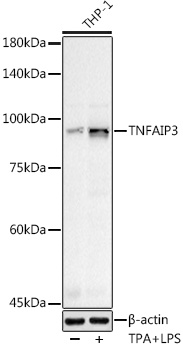
Western blot analysis of various lysates using (CAB2127) at 1:500 dilution. THP-1 cells were treated with PMA/TPA (200 nM) at 37℃ for 15 minutes after serum-starvation overnight. THP-1 cells were treated with LPS (1 μg/ml) at 37℃ for 8 hours. Secondary antibody: HRP-conjugated Goat anti-Rabbit IgG (H+L) (AS014) at 1:10000 dilution. Lysates/proteins: 25μg per lane. Blocking buffer: 3% nonfat dry milk in TBST. Detection: ECL Basic Kit (AbGn00020). Exposure time: 40s.
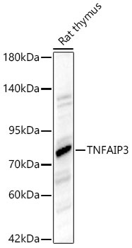
Western blot analysis of lysates from Rat thymus using TNFAIP3 Rabbit pAb (CAB2127) at 1:1000 dilution. Secondary antibody: HRP-conjugated Goat anti-Rabbit IgG (H+L) (AS014) at 1:10000 dilution. Lysates/proteins: 25 μg per lane. Blocking buffer: 3% nonfat dry milk in TBST. Detection: ECL Basic Kit (AbGn00020). Exposure time:20s.
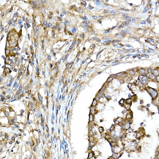
Immunohistochemistry analysis of paraffin-embedded Human lung cancer using TNFAIP3 Rabbit pAb (CAB2127) at dilution of 1:50 (40x lens). High pressure antigen retrieval performed with 0.01M Citrate buffer (pH 6.0) prior to IHC staining.
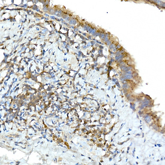
Immunohistochemistry analysis of paraffin-embedded Rat lung using TNFAIP3 Rabbit pAb (CAB2127) at dilution of 1:50 (40x lens). High pressure antigen retrieval performed with 0.01M Citrate buffer (pH 6.0) prior to IHC staining.


")


