The N-WASP/WASL Antibody (CAB2576) is a high-quality antibody developed for reliable detection and analysis of target proteins. This gene encodes a member of the Wiskott-Aldrich syndrome (WAS) protein family. Wiskott-Aldrich syndrome proteins share similar domain structure, and associate with a variety of signaling molecules to alter the actin cytoskeleton. The encoded protein is highly expressed in neural tissues, and interacts with several proteins involved in cytoskeletal organization, including cell division control protein 42 (CDC42) and the actin-related protein-2/3 (ARP2/3) complex. The encoded protein may be involved in the formation of long actin microspikes, and in neurite extension.
This antibody is validated for use in WB, IHC-P, IF/ICC, ELISA applications and has demonstrated reactivity against Human, Mouse, Rat samples.
Product Name:
N-WASP/WASL Antibody
SKU:
CAB2576
Size:
100μL, 20μL
Reactivity:
Human, Mouse, Rat
Conjugate:
Unconjugated
Immunogen:
Recombinant protein (or fragment).This information is considered to be commercially sensitive.
Tested Applications:
WBIHC-PIF/ICCELISA
Recommended Dilution:
WB
1:500 - 1:2000
IHC-P
1:50 - 1:200
IF/ICC
1:50 - 1:200
ELISA
Recommended starting concentration is 1 μg/mL. Please optimize the concentration based on your specific assay requirements.
Synonyms:
NWASP, WASPB, N-WASP, N-WASP/WASL
Positive Sample:
Rat kidney, HeLa, Mouse lung
Cellular Localization:
Cytoplasm, Nucleus, Cytoskeleton.
Calculated MW:
55kDa
Observed MW:
65kDa/
This gene encodes a member of the Wiskott-Aldrich syndrome (WAS) protein family. Wiskott-Aldrich syndrome proteins share similar domain structure, and associate with a variety of signaling molecules to alter the actin cytoskeleton. The encoded protein is highly expressed in neural tissues, and interacts with several proteins involved in cytoskeletal organization, including cell division control protein 42 (CDC42) and the actin-related protein-2/3 (ARP2/3) complex. The encoded protein may be involved in the formation of long actin microspikes, and in neurite extension.
Purification Method
Affinity purification
Gene ID
8976
RRID
AB_2764462
Buffer Information
Store at -20℃. Avoid freeze / thaw cycles. Buffer: PBS with 0.01% thimerosal,50% glycerol,pH7.3.
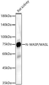
Western blot analysis of lysates from Rat kidney using N-WASP/WASL Rabbit pAb (CAB2576) at 1:2000 dilution. Secondary antibody: HRP-conjugated Goat anti-Rabbit IgG (H+L) (AS014) at 1:10000 dilution. Lysates/proteins: 25 μg per lane. Blocking buffer: 3% nonfat dry milk in TBST. Detection: ECL Basic Kit (AbGn00020). Exposure time: 30s.
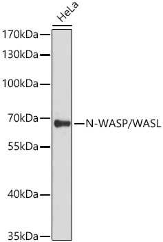
Western blot analysis of lysates from HeLa cells using N-WASP/WASL Rabbit pAb (CAB2576) at 1:3000 dilution incubated overnight at 4℃. Secondary antibody: HRP-conjugated Goat anti-Rabbit IgG (H+L) (AS014) at 1:10000 dilution. Lysates/proteins: 25 μg per lane. Blocking buffer: 3% nonfat dry milk in TBST. Detection: ECL Basic Kit (AbGn00020). Exposure time: 30s.
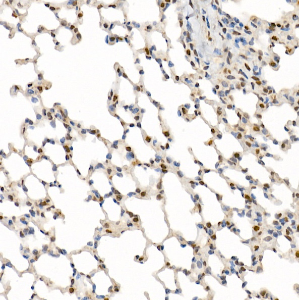
Immunohistochemistry analysis of paraffin-embedded Mouse lung using N-WASP/WASL Rabbit pAb (CAB2576) at dilution of 1:200 (40x lens). High pressure antigen retrieval performed with 0.01M Citrate buffer (pH 6.0) prior to IHC staining.
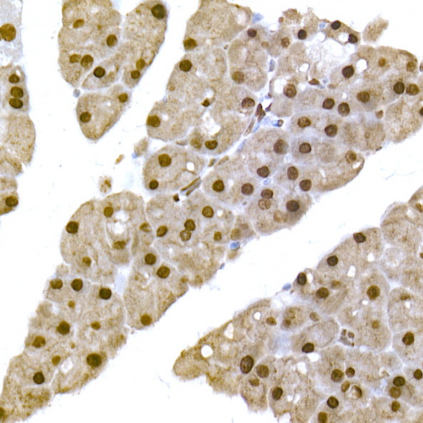
Immunohistochemistry analysis of paraffin-embedded Mouse pancreas using N-WASP/WASL Rabbit pAb (CAB2576) at dilution of 1:200 (40x lens). High pressure antigen retrieval performed with 0.01M Citrate buffer (pH 6.0) prior to IHC staining.
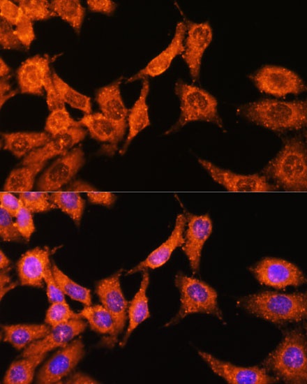
Immunofluorescence analysis of HeLa cells using N-WASP/WASL Rabbit pAb (CAB2576) at dilution of 1:100 (40x lens). Secondary antibody: Cy3-conjugated Goat anti-Rabbit IgG (H+L) (AS007) at 1:500 dilution. Blue: DAPI for nuclear staining.
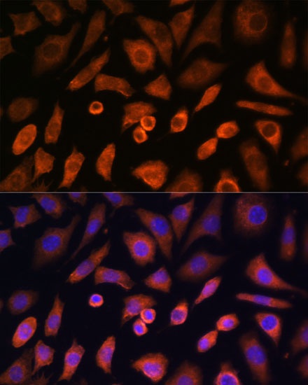
Immunofluorescence analysis of L-929 cells using N-WASP/WASL Rabbit pAb (CAB2576) at dilution of 1:100 (40x lens). Secondary antibody: Cy3-conjugated Goat anti-Rabbit IgG (H+L) (AS007) at 1:500 dilution. Blue: DAPI for nuclear staining.