The AARS Antibody (CAB15017) is a high-quality antibody developed for reliable detection and analysis of target proteins. This antibody, generated in rabbits, exhibits high reactivity with human samples and has been validated for use in Western blot applications. By binding specifically to the AARS protein, this antibody enables researchers to detect and analyze AARS expression in various cell types, making it an ideal choice for studies in molecular biology and biochemistry.AARS, also known as alanyl-tRNA synthetase, plays a crucial role in protein synthesis by catalyzing the attachment of alanine to transfer RNA molecules.
This antibody is validated for use in WB, IHC-P, IF/ICC, ELISA applications and has demonstrated reactivity against Human, Mouse, Rat samples.
Product Name:
AARS Antibody
SKU:
CAB15017
Size:
20μL, 100μL
Reactivity:
Human, Mouse, Rat
Conjugate:
Unconjugated
Immunogen:
Synthetic peptide. This information is considered to be commercially sensitive.
Recommended starting concentration is 1 μg/mL. Please optimize the concentration based on your specific assay requirements.
Synonyms:
AARS, TTD8, CMT2N, DEE29, HDLS2, EIEE29
Positive Sample:
HeLa, Jurkat, Mouse brain, Rat liver
Cellular Localization:
Cytoplasm.
Calculated MW:
107kDa
Observed MW:
107kDa
The human alanyl-tRNA synthetase (AARS) belongs to a family of tRNA synthases, of the class II enzymes. Class II tRNA synthases evolved early in evolution and are highly conserved. This is reflected by the fact that 498 of the 968-residue polypeptide human AARS shares 41% identity witht the E.coli protein. tRNA synthases are the enzymes that interpret the RNA code and attach specific aminoacids to the tRNAs that contain the cognate trinucleotide anticodons. They consist of a catalytic domain which interacts with the amino acid acceptor-T psi C helix of the tRNA, and a second domain which interacts with the rest of the tRNA structure.
Purification Method
Affinity purification
Gene ID
16
RRID
AB_2761897
Buffer Information
Store at -20℃. Avoid freeze / thaw cycles. Buffer: PBS with 0.09% sodium azide,50% glycerol,pH7.3.
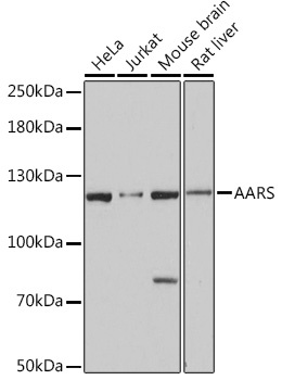
Western blot analysis of various lysates using AARS Rabbit pAb (CAB15017) at 1:3000 dilution. Secondary antibody: HRP-conjugated Goat anti-Rabbit IgG (H+L) (CABS014) at 1:10000 dilution. Lysates/proteins: 25μg per lane. Blocking buffer: 3% nonfat dry milk in TBST. Detection: ECL Basic Kit (AbGn00020). Exposure time: 10s.
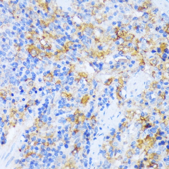
Immunohistochemistry analysis of paraffin-embedded Rat spleen using AARS Rabbit pAb (CAB15017) at dilution of 1:100 (40x lens). Microwave antigen retrieval performed with 0.01M PBS Buffer (pH 7.2) prior to IHC staining.
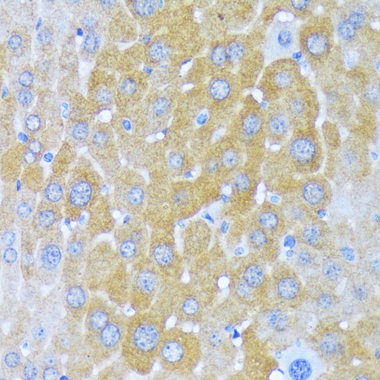
Immunohistochemistry analysis of paraffin-embedded Mouse liver using AARS Rabbit pAb (CAB15017) at dilution of 1:100 (40x lens). Microwave antigen retrieval performed with 0.01M PBS Buffer (pH 7.2) prior to IHC staining.
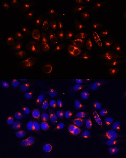
Immunofluorescence analysis of 293T cells using AARS Rabbit pAb (CAB15017) at dilution of 1:100 (40x lens). Secondary antibody: Cy3-conjugated Goat anti-Rabbit IgG (H+L) (CABS007) at 1:500 dilution. Blue: DAPI for nuclear staining.