The ABHD4 Antibody (CAB17751) is a high-quality antibody developed for reliable detection and analysis of target proteins. This antibody, developed in rabbits, exhibits high reactivity with human samples and has been validated for use in Western blot assays. By targeting the ABHD4 protein, this antibody allows for the detection and analysis of ABHD4 expression in various cell types, making it a useful tool for investigations in biochemistry, cell biology, and lipid metabolism research.
This antibody is validated for use in WB, IF/ICC, ELISA applications and has demonstrated reactivity against Human, Mouse, Rat samples.
Product Name:
ABHD4 Antibody
SKU:
CAB17751
Size:
20μL, 100μL
Reactivity:
Human, Mouse, Rat
Conjugate:
Unconjugated
Immunogen:
Recombinant protein (or fragment).This information is considered to be commercially sensitive.
Recommended starting concentration is 1 μg/mL. Please optimize the concentration based on your specific assay requirements.
Synonyms:
ABH4, ABHD4
Positive Sample:
Rat testis
Cellular Localization:
Lipid Droplet, Mitochondrion.
Calculated MW:
39kDa
Observed MW:
39kDa
Predicted to enable lysophosphatidic acid acyltransferase activity and lysophospholipase activity. Predicted to be involved in N-acylphosphatidylethanolamine metabolic process; lipid homeostasis; and phosphatidic acid biosynthetic process. Predicted to act upstream of or within N-acylethanolamine metabolic process. Predicted to be located in endoplasmic reticulum membrane. Predicted to be active in lipid droplet and mitochondrion.
Purification Method
Affinity purification
Gene ID
63874
RRID
AB_2768188
Buffer Information
Store at -20℃. Avoid freeze / thaw cycles. Buffer: PBS with 0.01% thimerosal,50% glycerol,pH7.3.
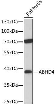
Western blot analysis of lysates from Rat testis, using ABHD4 Rabbit pAb (CAB17751) at 1:1000 dilution. Secondary antibody: HRP-conjugated Goat anti-Rabbit IgG (H+L) (CABS014) at 1:10000 dilution. Lysates/proteins: 25μg per lane. Blocking buffer: 3% nonfat dry milk in TBST. Detection: ECL Basic Kit (AbGn00020). Exposure time: 30s.
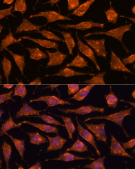
Immunofluorescence analysis of L929 cells using ABHD4 Rabbit pAb (CAB17751) at dilution of 1:100. Secondary antibody: Cy3-conjugated Goat anti-Rabbit IgG (H+L) (CABS007) at 1:500 dilution. Blue: DAPI for nuclear staining.