The ACAA2 Antibody (CAB15778) is a high-quality antibody developed for reliable detection and analysis of target proteins. The encoded protein catalyzes the last step of the mitochondrial fatty acid beta-oxidation spiral. Unlike most mitochondrial matrix proteins, it contains a non-cleavable amino-terminal targeting signal.
This antibody is validated for use in WB, IHC-P, IF/ICC, ELISA applications and has demonstrated reactivity against Human, Mouse, Rat samples.
Product Name:
ACAA2 Antibody
SKU:
CAB15778
Size:
100μL, 20μL
Reactivity:
Human, Mouse, Rat
Conjugate:
Unconjugated
Immunogen:
Recombinant protein (or fragment).This information is considered to be commercially sensitive.
Tested Applications:
WBIHC-PIF/ICCELISA
Recommended Dilution:
WB
1:500 - 1:2000
IHC-P
1:50 - 1:200
IF/ICC
1:50 - 1:200
ELISA
Recommended starting concentration is 1 μg/mL. Please optimize the concentration based on your specific assay requirements.
Synonyms:
DSAEC, ACAA2
Positive Sample:
Hep G2, HeLa, Mouse liver
Cellular Localization:
Mitochondrion.
Calculated MW:
42kDa
Observed MW:
42kDa
The encoded protein catalyzes the last step of the mitochondrial fatty acid beta-oxidation spiral. Unlike most mitochondrial matrix proteins, it contains a non-cleavable amino-terminal targeting signal.
Purification Method
Affinity purification
Gene ID
10449
RRID
AB_2763197
Buffer Information
Store at -20℃. Avoid freeze / thaw cycles. Buffer: PBS containing 50% glycerol, preserved with proclin300 or sodium azide, pH 7.3.
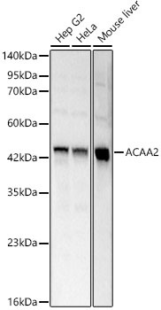
Western blot analysis of various lysates using ACAA2 Rabbit pAb (CAB15778) at 1:1000 dilution. Secondary antibody: HRP-conjugated Goat anti-Rabbit IgG (H+L) (AS014) at 1:10000 dilution. Lysates / proteins: 25 μg per lane. Blocking buffer: 3 % nonfat dry milk in TBST. Detection: ECL Basic Kit (AbGn00020). Exposure time: 0.8s.
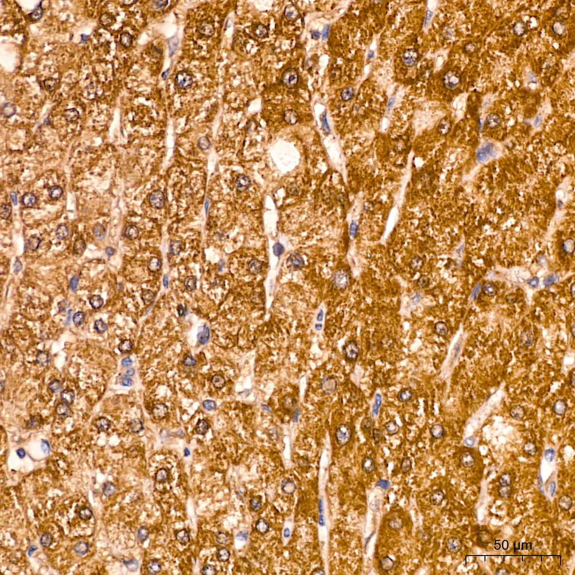
Immunohistochemistry analysis of paraffin-embedded Human liver tissue using ACAA2 Rabbit pAb (CAB15778) at a dilution of 1:100 (40x lens). High pressure antigen retrieval was performed with 0.01 M citrate buffer (pH 6.0) prior to IHC staining.
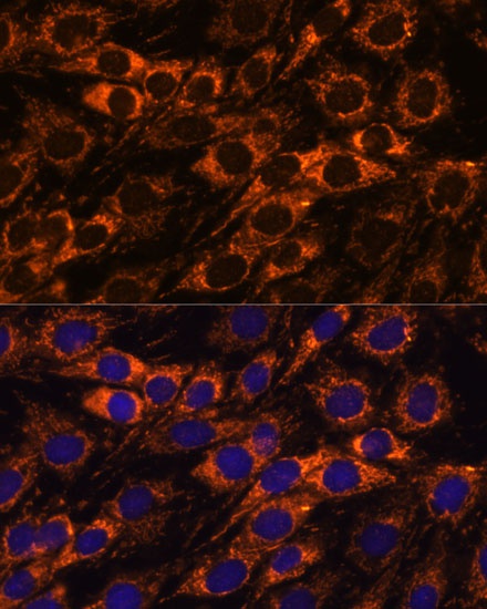
Immunofluorescence analysis of C6 cells using ACAA2 Rabbit pAb (CAB15778) at dilution of 1:100. Secondary antibody: Cy3-conjugated Goat anti-Rabbit IgG (H+L) (AS007) at 1:500 dilution. Blue: DAPI for nuclear staining.
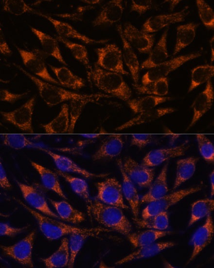
Immunofluorescence analysis of L929 cells using ACAA2 Rabbit pAb (CAB15778) at dilution of 1:100. Secondary antibody: Cy3-conjugated Goat anti-Rabbit IgG (H+L) (AS007) at 1:500 dilution. Blue: DAPI for nuclear staining.