The acetyl-ACC1 Antibody (CAB15606) is a high-quality antibody developed for reliable detection and analysis of target proteins. Raised in rabbits, this antibody is highly specific to human samples and has been validated for use in Western blot applications. By binding to the ACACA protein, this antibody allows for the precise detection and analysis of ACACA in a variety of cell types, making it an essential component for studies in metabolism and related research areas.ACACA, also known as acetyl-CoA carboxylase alpha, plays a crucial role in fatty acid synthesis and energy regulation.
This antibody is validated for use in WB, IHC-P, IF/ICC, IP, ELISA applications and has demonstrated reactivity against Human, Mouse, Rat samples.
Product Name:
acetyl-ACC1 Antibody
SKU:
CAB15606
Size:
20μL, 100μL
Reactivity:
Human, Mouse, Rat
Conjugate:
Unconjugated
Immunogen:
Recombinant protein (or fragment).This information is considered to be commercially sensitive.
Acetyl-CoA carboxylase (ACC) is a complex multifunctional enzyme system. ACC is a biotin-containing enzyme which catalyzes the carboxylation of acetyl-CoA to malonyl-CoA, the rate-limiting step in fatty acid synthesis. There are two ACC forms, alpha and beta, encoded by two different genes. ACC-alpha is highly enriched in lipogenic tissues. The enzyme is under long term control at the transcriptional and translational levels and under short term regulation by the phosphorylation/dephosphorylation of targeted serine residues and by allosteric transformation by citrate or palmitoyl-CoA. Multiple alternatively spliced transcript variants divergent in the 5' sequence and encoding distinct isoforms have been found for this gene.
Purification Method
Affinity purification
Gene ID
31
RRID
AB_2763012
Buffer Information
Store at -20℃. Avoid freeze / thaw cycles. Buffer: PBS containing 50% glycerol, preserved with proclin300 or sodium azide, pH 7.3.
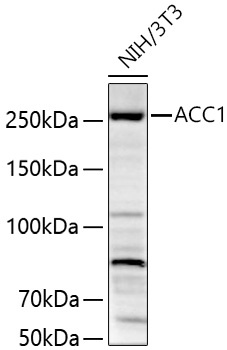
Western blot analysis of lysates from NIH/3T3 cells using ACC1 Rabbit pAb (CAB15606) at 1:1000 dilution incubated overnight at 4℃. Secondary antibody: HRP-conjugated Goat anti-Rabbit IgG (H+L) (CABS014) at 1:10000 dilution. Lysates/proteins: 25 μg per lane. Blocking buffer: 3% nonfat dry milk in TBST. Detection: ECL Basic Kit (AbGn00020). Exposure time: 45 s.
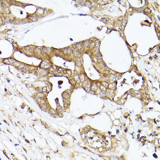
Immunohistochemistry analysis of paraffin-embedded Human colon carcinoma using ACC1 Rabbit pAb (CAB15606) at dilution of 1:50 (40x lens). High pressure antigen retrieval performed with 0.01M Citrate buffer (pH 6.0) prior to IHC staining.
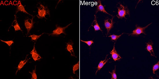
Immunofluorescence analysis of C6 cells using ACC1 Rabbit pAb (CAB15606) at dilution of 1:100 (40x lens). Secondary antibody: Cy3-conjugated Goat anti-Rabbit IgG (H+L) (CABS007) at 1:500 dilution. Blue: DAPI for nuclear staining.
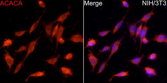
Immunofluorescence analysis of NIH/3T3 cells using ACC1 Rabbit pAb (CAB15606) at dilution of 1:100 (40x lens). Secondary antibody: Cy3-conjugated Goat anti-Rabbit IgG (H+L) (CABS007) at 1:500 dilution. Blue: DAPI for nuclear staining.