The ACADS Antibody (CAB0945) is a high-quality antibody developed for reliable detection and analysis of target proteins. This antibody, raised in rabbits, is highly specific to human ACADS and has been validated for use in Western blot applications. By binding to the ACADS protein, this antibody enables the detection and analysis of ACADS in various cell types, making it an essential component of studies in metabolic disorders, lipid metabolism, and related diseases.ACADS plays a crucial role in the breakdown of fatty acids for energy production, making it a target of interest in research on metabolic diseases such as fatty acid oxidation disorders and mitochondrial dysfunction.
This antibody is validated for use in WB, IHC-P, ELISA applications and has demonstrated reactivity against Human, Mouse, Rat samples.
Product Name:
ACADS Antibody
SKU:
CAB0945
Size:
20μL, 100μL
Reactivity:
Human, Mouse, Rat
Conjugate:
Unconjugated
Immunogen:
Recombinant protein (or fragment).This information is considered to be commercially sensitive.
Recommended starting concentration is 1 μg/mL. Please optimize the concentration based on your specific assay requirements.
Synonyms:
SCAD, ACAD3, ACADS
Positive Sample:
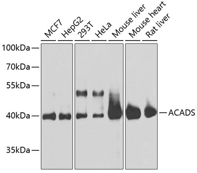
MCF7, HepG2, 293T, HeLa, Mouse liver, Mouse heart, Rat liver
Cellular Localization:
Mitochondrion Matrix.
Calculated MW:
44kDa
Observed MW:
44kDa
This gene encodes a tetrameric mitochondrial flavoprotein, which is a member of the acyl-CoA dehydrogenase family. This enzyme catalyzes the initial step of the mitochondrial fatty acid beta-oxidation pathway. Mutations in this gene have been associated with short-chain acyl-CoA dehydrogenase (SCAD) deficiency. Alternative splicing results in two variants which encode different isoforms.
Purification Method
Affinity purification
Gene ID
35
RRID
AB_2757471
Buffer Information
Store at -20℃. Avoid freeze / thaw cycles. Buffer: PBS containing 50% glycerol, preserved with proclin300 or sodium azide, pH 7.3.
Western blot analysis of various lysates using ACADS Rabbit pAb (CAB0945) at 1:1000 dilution. Secondary antibody: HRP-conjugated Goat anti-Rabbit IgG (H+L) (CABS014) at 1:10000 dilution. Lysates/proteins: 25μg per lane. Blocking buffer: 3% nonfat dry milk in TBST. Detection: ECL Basic Kit (AbGn00020). Exposure time: 15s.
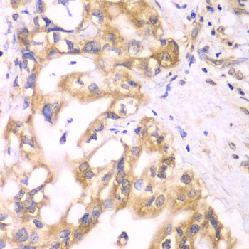
Immunohistochemistry analysis of paraffin-embedded Human liver cancer using ACADS Rabbit pAb (CAB0945) at dilution of 1:100 (40x lens). Microwave antigen retrieval performed with 0.01M PBS Buffer (pH 7.2) prior to IHC staining.