The ACSL4 Antibody (CAB6826) is a high-quality antibody developed for reliable detection and analysis of target proteins. This antibody, generated in rabbits, exhibits high reactivity with human samples and has been validated for use in Western blot applications. By specifically binding to ACSL4, this antibody enables accurate detection and analysis of the enzyme in different cell types, making it ideal for investigations into lipid metabolism, cancer biology, and neurological diseases.
This antibody is validated for use in WB, IHC-P, IF/ICC, IP, ELISA applications and has demonstrated reactivity against Human, Mouse, Rat samples.
Product Name:
ACSL4 Antibody
SKU:
CAB6826
Size:
20μL, 100μL
Reactivity:
Human, Mouse, Rat
Conjugate:
Unconjugated
Immunogen:
Recombinant protein (or fragment).This information is considered to be commercially sensitive.
0.5μg-4μg antibody for 200μg-400μg extracts of whole cells
IF/ICC
1:50 - 1:200
IHC-P
1:50 - 1:200
ELISA
Recommended starting concentration is 1 μg/mL. Please optimize the concentration based on your specific assay requirements.
Synonyms:
ACS4, FACL4, LACS4, MRX63, MRX68, XLID63, ACSL4
Positive Sample:
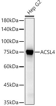
Hep G2
Cellular Localization:
Endoplasmic Reticulum Membrane, Microsome Membrane, Mitochondrion Outer Membrane, Peroxisome Membrane, Single-Pass Type Iii Membrane Protein.
Calculated MW:
79kDa
Observed MW:
79kDa
The protein encoded by this gene is an isozyme of the long-chain fatty-acid-coenzyme A ligase family. Although differing in substrate specificity, subcellular localization, and tissue distribution, all isozymes of this family convert free long-chain fatty acids into fatty acyl-CoA esters, and thereby play a key role in lipid biosynthesis and fatty acid degradation. This isozyme preferentially utilizes arachidonate as substrate. The absence of this enzyme may contribute to the cognitive disability or Alport syndrome. Alternative splicing of this gene generates multiple transcript variants.
Purification Method
Affinity purification
Gene ID
2182
RRID
AB_2767400
Buffer Information
Store at -20℃. Avoid freeze / thaw cycles. Buffer: PBS containing 50% glycerol, preserved with proclin300 or sodium azide, pH 7.3.
Western blot analysis of lysates from Hep G2 cells using ACSL4 Rabbit pAb (CAB6826) at 1:400 dilution. Secondary antibody: HRP-conjugated Goat anti-Rabbit IgG (H+L) (CABS014) at 1:10000 dilution. Lysates/proteins: 25 μg per lane. Blocking buffer: 3% nonfat dry milk in TBST. Detection: ECL Basic Kit (AbGn00020). Exposure time: 10s.
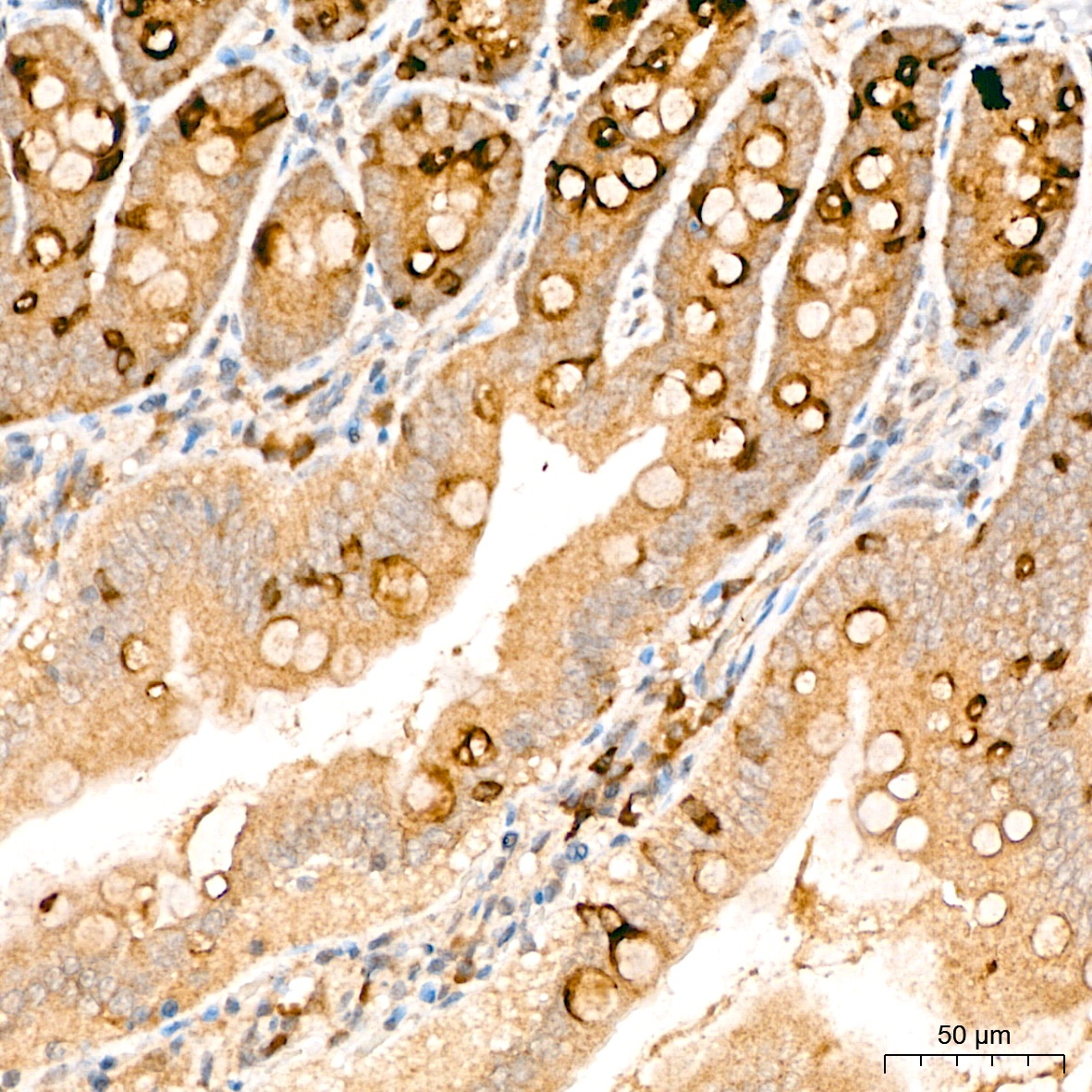
Immunohistochemistry analysis of paraffin-embedded Rat colon tissue using ACSL4 Rabbit pAb (CAB6826) at a dilution of 1:200 (40x lens). High pressure antigen retrieval performed with 0.01M Citrate Buffer(pH 6.0) prior to IHC staining.
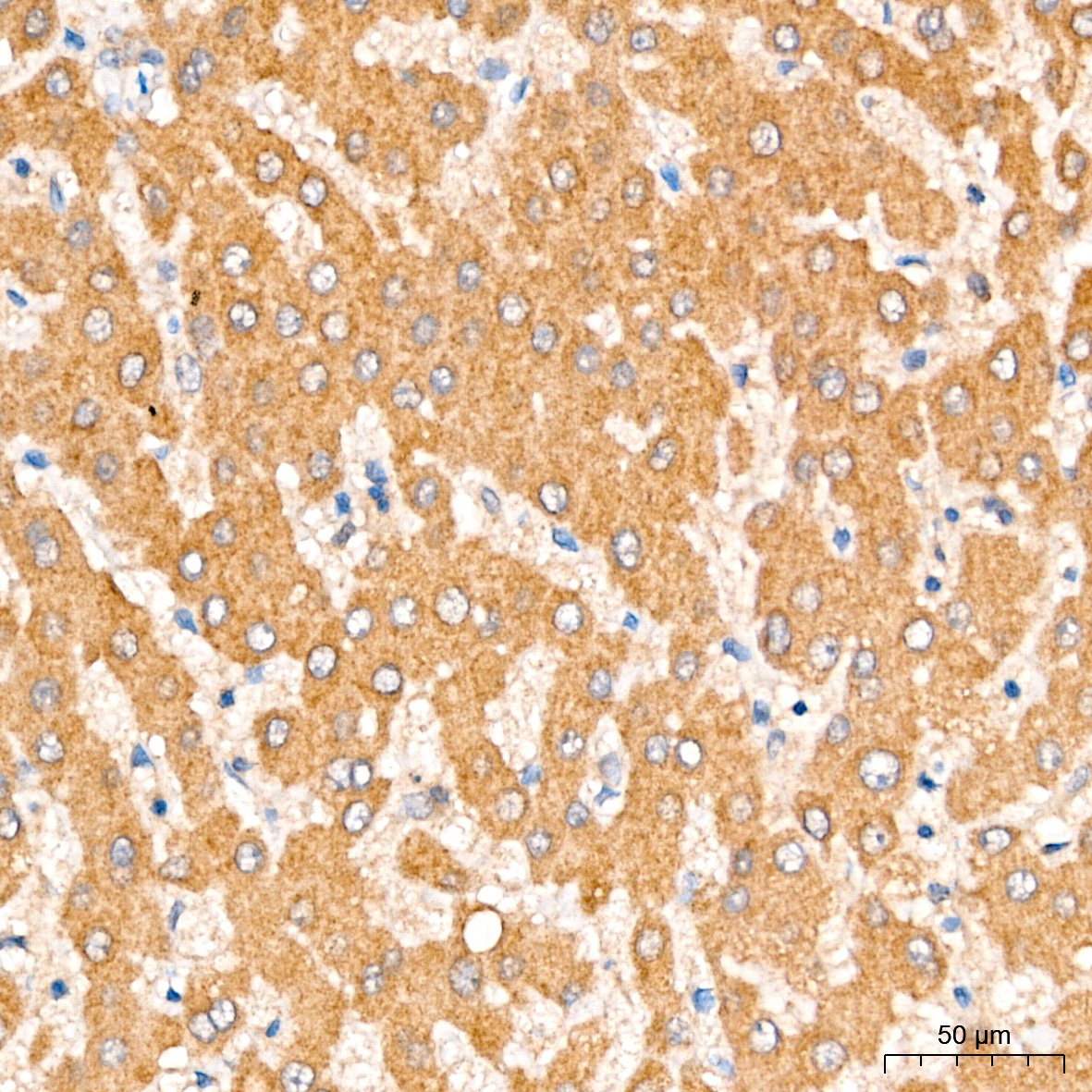
Immunohistochemistry analysis of paraffin-embedded Human liver tissue using ACSL4 Rabbit pAb (CAB6826) at a dilution of 1:200 (40x lens). High pressure antigen retrieval performed with 0.01M Citrate Buffer(pH 6.0) prior to IHC staining.
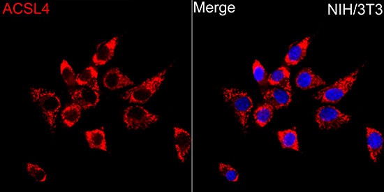
Immunofluorescence analysis of NIH/3T3 cells using ACSL4 Rabbit pAb (CAB6826) at dilution of 1:50 (40x lens). Secondary antibody: Cy3-conjugated Goat anti-Rabbit IgG (H+L) (CABS007) at 1:500 dilution. Blue: DAPI for nuclear staining.