The ACTC1 Antibody (CAB6507) is a high-quality antibody developed for reliable detection and analysis of target proteins. This polyclonal antibody, raised in rabbits, is validated for use in various applications, including Western blot and immunohistochemistry.ACTC1, also known as cardiac muscle alpha actin, is primarily expressed in cardiac muscle cells and is essential for maintaining the structural integrity of the sarcomere. Dysregulation of ACTC1 has been linked to various cardiac disorders, making it a crucial target for research in cardiology and muscle biology.
This antibody is validated for use in WB, IHC-P, ELISA applications and has demonstrated reactivity against Human, Mouse, Rat samples.
Product Name:
ACTC1 Antibody
SKU:
CAB6507
Size:
20μL, 100μL
Reactivity:
Human, Mouse, Rat
Conjugate:
Unconjugated
Immunogen:
Synthetic peptide. This information is considered to be commercially sensitive.
Recommended starting concentration is 1 μg/mL. Please optimize the concentration based on your specific assay requirements.
Synonyms:
ACTC, ASD5, CMD1R, CMH11, LVNC4, ACTC1
Positive Sample:
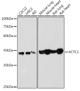
C2C12, H9C2, RD, Mouse lung, Mouse heart, Rat lung, Rat heart
Cellular Localization:
Cytoplasm, Cytoskeleton.
Calculated MW:
42kDa
Observed MW:
42kDa
Actins are highly conserved proteins that are involved in various types of cell motility. Polymerization of globular actin (G-actin) leads to a structural filament (F-actin) in the form of a two-stranded helix. Each actin can bind to four others. The protein encoded by this gene belongs to the actin family which is comprised of three main groups of actin isoforms, alpha, beta, and gamma. The alpha actins are found in muscle tissues and are a major constituent of the contractile apparatus. Defects in this gene have been associated with idiopathic dilated cardiomyopathy (IDC) and familial hypertrophic cardiomyopathy (FHC).
Purification Method
Affinity purification
Gene ID
70
RRID
AB_2767103
Buffer Information
Store at -20℃. Avoid freeze / thaw cycles. Buffer: PBS containing 50% glycerol, preserved with proclin300 or sodium azide, pH 7.3.
Western blot analysis of various lysates using ACTC1 Rabbit pAb (CAB6507) at 1:1000 dilution. Secondary antibody: HRP-conjugated Goat anti-Rabbit IgG (H+L) (CABS014) at 1:10000 dilution. Lysates/proteins: 25μg per lane. Blocking buffer: 3% nonfat dry milk in TBST. Detection: ECL Basic Kit (AbGn00020). Exposure time: 10s.
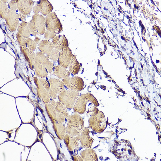
Immunohistochemistry analysis of paraffin-embedded Human skeletal muscle using ACTC1 Rabbit pAb (CAB6507) at dilution of 1:50 (40x lens). High pressure antigen retrieval performed with 0.01M Citrate buffer (pH 6.0) prior to IHC staining.