The EMR1 Antibody (CAB1256) is a high-quality antibody developed for reliable detection and analysis of target proteins. This antibody, generated in rabbits, exhibits high reactivity towards human samples and has been validated for use in Western blot applications. By specifically binding to the EMR1 protein, researchers can accurately detect and analyze its expression in various cell types, making it an invaluable tool for investigations in immunology and cancer research.EMR1, also known as EGF-like module-containing mucin-like hormone receptor-like 1, plays a crucial role in immune modulation by regulating inflammatory responses and potentially influencing allergic reactions.
This antibody is validated for use in WB, IF/ICC, ELISA, IF-P applications and has demonstrated reactivity against Human, Mouse, Rat samples.
Product Name:
EMR1 Antibody
SKU:
CAB1256
Size:
20μL, 100μL
Reactivity:
Human, Mouse, Rat
Conjugate:
Unconjugated
Immunogen:
Recombinant protein (or fragment).This information is considered to be commercially sensitive.
Recommended starting concentration is 1 μg/mL. Please optimize the concentration based on your specific assay requirements.
Synonyms:
EMR1, TM7LN3
Positive Sample:
Mouse lung
Cellular Localization:
Cell Membrane, Multi-Pass Membrane Protein.
Calculated MW:
98kDa
Observed MW:
160kDa
This gene encodes a protein that has a domain resembling seven transmembrane G protein-coupled hormone receptors (7TM receptors) at its C-terminus. The N-terminus of the encoded protein has six EGF-like modules, separated from the transmembrane segments by a serine/threonine-rich domain, a feature reminiscent of mucin-like, single-span, integral membrane glycoproteins with adhesive properties. Multiple alternatively spliced transcript variants encoding different isoforms have been found for this gene.
Purification Method
Affinity purification
Gene ID
2015
RRID
AB_2759401
Buffer Information
Store at -20℃. Avoid freeze / thaw cycles. Buffer: PBS containing 50% glycerol, preserved with proclin300 or sodium azide, pH 7.3.
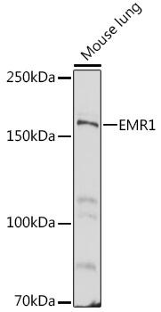
Western blot analysis of lysates from Mouse lung, using EMR1 Rabbit pAb (CAB1256) at 1:500 dilution. Secondary antibody: HRP-conjugated Goat anti-Rabbit IgG (H+L) (CABS014) at 1:10000 dilution. Lysates/proteins: 25μg per lane. Blocking buffer: 3% nonfat dry milk in TBST. Detection: ECL Basic Kit (AbGn00020). Exposure time: 60s.
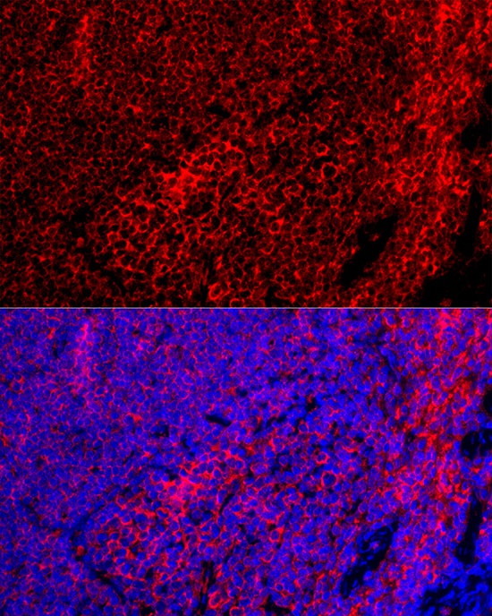
Immunofluorescence analysis of paraffin-embedded Mouse spleen using EMR1 Rabbit pAb (CAB1256) at dilution of 1:100 (40x lens). Secondary antibody: Cy3-conjugated Goat anti-Rabbit IgG (H+L) (CABS007) at 1:500 dilution. Blue: DAPI for nuclear staining.
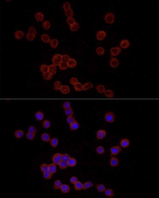
Immunofluorescence analysis of THP-1 cells using EMR1 Rabbit pAb (CAB1256) at dilution of 1:25 (40x lens). Secondary antibody: Cy3-conjugated Goat anti-Rabbit IgG (H+L) (CABS007) at 1:500 dilution. Blue: DAPI for nuclear staining.