The ADH1A/ADH1B/ADH1C Antibody (CAB18581) is a high-quality antibody developed for reliable detection and analysis of target proteins. These enzymes play a crucial role in alcohol metabolism, converting alcohol into acetaldehyde in the liver. The antibody, raised in rabbits, is highly specific and reactive with human samples, making it a reliable choice for immunohistochemistry and Western blot applications.ADH1A, ADH1B, and ADH1C are key players in ethanol metabolism, with variations in these genes affecting an individual's alcohol tolerance and susceptibility to alcohol-related diseases.
This antibody is validated for use in WB, ELISA, IF-P applications and has demonstrated reactivity against Human, Mouse, Rat samples.
Product Name:
ADH1A/ADH1B/ADH1C Antibody
SKU:
CAB18581
Size:
20μL, 100μL
Reactivity:
Human, Mouse, Rat
Immunogen:
Synthetic peptide. This information is considered to be commercially sensitive.
Recommended starting concentration is 1 μg/mL. Please optimize the concentration based on your specific assay requirements.
Synonyms:
ADH1A/ADH1B/ADH1C
Positive Sample:
Mouse lung
Cellular Localization:
Cytosol, Nucleoplasm, Plasma Membrane.
Observed MW:
40kDa
Alcohol dehydrogenase,consisting of several homo- and heterodimers of alpha, beta, and gamma subunits, exhibits high activity for ethanol oxidation and plays a major role in ethanol catabolism.Members of this enzyme family metabolize a wide variety of substrates, including ethanol, retinol, other aliphatic alcohols, hydroxysteroids, and lipid peroxidation products. Three genes encoding alpha, beta and gamma subunits are tandemly organized in a genomic segment as a gene cluster.
Purification Method
Affinity purification
Gene ID
124 125 126
RRID
AB_2862341
Buffer Information
Store at -20℃. Avoid freeze / thaw cycles. Buffer: PBS with 0.01% thimerosal,50% glycerol,pH7.3.
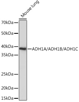
Western blot analysis of lysates from Mouse lung, using ADH1A/ADH1B/ADH1C Rabbit pAb (CAB18581) at 1:1000 dilution. Secondary antibody: HRP-conjugated Goat anti-Rabbit IgG (H+L) (CABS014) at 1:10000 dilution. Lysates/proteins: 25μg per lane. Blocking buffer: 3% nonfat dry milk in TBST. Detection: ECL Basic Kit (AbGn00020). Exposure time: 10s.
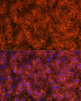
Immunofluorescence analysis of paraffin-embedded Human liver using ADH1A/ADH1B/ADH1C Rabbit pAb (CAB18581) at dilution of 1:100. Secondary antibody: Cy3-conjugated Goat anti-Rabbit IgG (H+L) (CABS007) at 1:500 dilution. Blue: DAPI for nuclear staining.
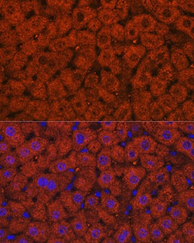
Immunofluorescence analysis of paraffin-embedded Mouse liver using ADH1A/ADH1B/ADH1C Rabbit pAb (CAB18581) at dilution of 1:100. Secondary antibody: Cy3-conjugated Goat anti-Rabbit IgG (H+L) (CABS007) at 1:500 dilution. Blue: DAPI for nuclear staining.