The ADH7 Antibody (CAB7871) is a high-quality antibody developed for reliable detection and analysis of target proteins. This antibody, produced in rabbits, exhibits high reactivity with human samples and has been validated for use in various applications, including Western blotting.ADH7 plays a crucial role in the breakdown of ethanol and retinol in the body, making it a key player in alcohol metabolism and vitamin A metabolism. Dysregulation of ADH7 has been linked to various health conditions, including alcohol dependence and vitamin A deficiency disorders.
This antibody is validated for use in WB, IF/ICC, ELISA applications and has demonstrated reactivity against Human, Mouse, Rat samples.
Product Name:
ADH7 Antibody
SKU:
CAB7871
Size:
20μL, 100μL
Reactivity:
Human, Mouse, Rat
Conjugate:
Unconjugated
Immunogen:
Recombinant protein (or fragment).This information is considered to be commercially sensitive.
Recommended starting concentration is 1 μg/mL. Please optimize the concentration based on your specific assay requirements.
Synonyms:
ADH4, ADH7
Positive Sample:
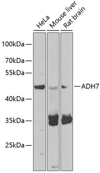
HeLa, Mouse liver, Rat brain
Cellular Localization:
Cytoplasm.
Calculated MW:
41kDa
Observed MW:
50kDa
This gene encodes class IV alcohol dehydrogenase 7 mu or sigma subunit, which is a member of the alcohol dehydrogenase family. Members of this family metabolize a wide variety of substrates, including ethanol, retinol, other aliphatic alcohols, hydroxysteroids, and lipid peroxidation products. The enzyme encoded by this gene is inefficient in ethanol oxidation, but is the most active as a retinol dehydrogenase; thus it may participate in the synthesis of retinoic acid, a hormone important for cellular differentiation. The expression of this gene is much more abundant in stomach than liver, thus differing from the other known gene family members. Alternative splicing results in multiple transcript variants.
Purification Method
Affinity purification
Gene ID
131
RRID
AB_2768261
Buffer Information
Store at -20℃. Avoid freeze / thaw cycles. Buffer: PBS containing 50% glycerol, preserved with proclin300 or sodium azide, pH 7.3.
Western blot analysis of various lysates using ADH7 Rabbit pAb (CAB7871) at 1:1000 dilution. Secondary antibody: HRP-conjugated Goat anti-Rabbit IgG (H+L) (CABS014) at 1:10000 dilution. Lysates/proteins: 25μg per lane. Blocking buffer: 3% nonfat dry milk in TBST. Detection: ECL Basic Kit (AbGn00020). Exposure time: 5s.
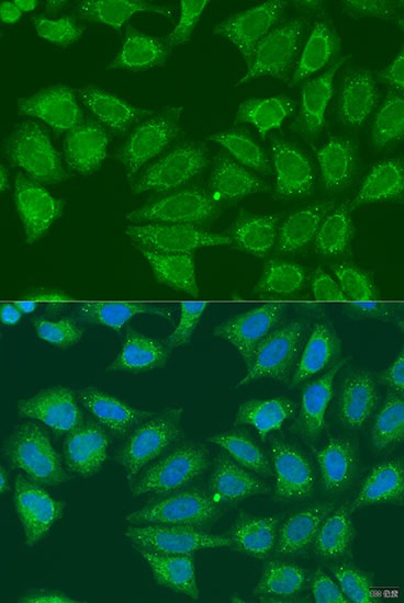
Immunofluorescence analysis of U2OS cells using ADH7 Rabbit pAb (CAB7871) at dilution of 1:100. Secondary antibody: Cy3-conjugated Goat anti-Rabbit IgG (H+L) (CABS007) at 1:500 dilution. Blue: DAPI for nuclear staining.