The AGER Antibody (CAB1395) is a high-quality antibody developed for reliable detection and analysis of target proteins. RAGE is involved in various physiological and pathological processes, including inflammation, cancer, diabetes, and neurodegenerative diseases. This antibody, produced in rabbits, specifically recognizes human AGER protein and is validated for use in Western blot applications. By binding to the AGER receptor, the antibody allows for the detection and analysis of AGER expression in different cell types, making it suitable for investigations in immunology, oncology, and neurobiology.
This antibody is validated for use in WB, ELISA applications and has demonstrated reactivity against Human, Mouse, Rat samples.
Product Name:
AGER Antibody
SKU:
CAB1395
Size:
20μL, 100μL
Reactivity:
Human, Mouse, Rat
Conjugate:
Unconjugated
Immunogen:
Recombinant protein (or fragment).This information is considered to be commercially sensitive.
Recommended starting concentration is 1 μg/mL. Please optimize the concentration based on your specific assay requirements.
Synonyms:
RAGE, sRAGE, SCARJ1, AGER
Positive Sample:
A-549, NCI-H460, Mouse small intestine, Rat kidney
Cellular Localization:
Cell Membrane, Secreted, Single-Pass Type I Membrane Protein.
Calculated MW:
43kDa
Observed MW:
43kDa
The advanced glycosylation end product (AGE) receptor encoded by this gene is a member of the immunoglobulin superfamily of cell surface receptors. It is a multiligand receptor, and besides AGE, interacts with other molecules implicated in homeostasis, development, and inflammation, and certain diseases, such as diabetes and Alzheimer's disease. Many alternatively spliced transcript variants encoding different isoforms, as well as non-protein-coding variants, have been described for this gene (PMID:18089847).
Purification Method
Affinity purification
Gene ID
177
RRID
AB_2760803
Buffer Information
Store at -20℃. Avoid freeze / thaw cycles. Buffer: PBS containing 50% glycerol, preserved with proclin300 or sodium azide, pH 7.3.
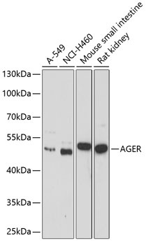
Western blot analysis of various lysates using AGER Rabbit pAb (CAB1395) at 1:1000 dilution. Secondary antibody: HRP-conjugated Goat anti-Rabbit IgG (H+L) (CABS014) at 1:10000 dilution. Lysates/proteins: 25μg per lane. Blocking buffer: 3% nonfat dry milk in TBST. Detection: ECL Basic Kit (AbGn00020). Exposure time: 90s.


 at 1:10000 dilution.
Lysates/proteins: 25ug per lane.
Blocking buffer: 3% nonfat dry milk in TBST.
Detection: ECL Basic Kit (RM00020).
Exposure time: 1s.")
")


