The AGK Antibody (CAB9976) is a high-quality antibody developed for reliable detection and analysis of target proteins. This antibody is produced in rabbits and shows high reactivity with human samples, making it a reliable choice for Western blot applications. By binding specifically to the AGK protein, this antibody enables the detection and analysis of AGK in various cell types, providing valuable insights into its function in metabolic processes and signaling pathways.
This antibody is validated for use in WB, ELISA applications and has demonstrated reactivity against Human, Rat samples.
Product Name:
AGK Antibody
SKU:
CAB9976
Size:
20μL, 100μL
Reactivity:
Human, Rat
Conjugate:
Unconjugated
Immunogen:
Recombinant protein (or fragment).This information is considered to be commercially sensitive.
Recommended starting concentration is 1 μg/mL. Please optimize the concentration based on your specific assay requirements.
Synonyms:
MULK, CATC5, CTRCT38, MTDPS10, AGK
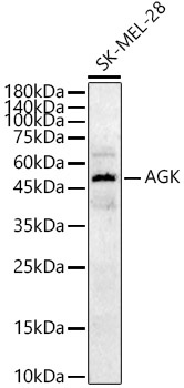
Positive Sample:
SK-MEL-28
Cellular Localization:
Mitochondrion Membrane.
Calculated MW:
47kDa
Observed MW:
47kDa
The protein encoded by this gene is a mitochondrial membrane protein involved in lipid and glycerolipid metabolism. The encoded protein is a lipid kinase that catalyzes the formation of phosphatidic and lysophosphatidic acids. Defects in this gene have been associated with mitochondrial DNA depletion syndrome 10.
Purification Method
Affinity purification
Gene ID
55750
RRID
AB_2768281
Buffer Information
Store at -20℃. Avoid freeze / thaw cycles. Buffer: PBS containing 50% glycerol, preserved with proclin300 or sodium azide, pH 7.3.
Western blot analysis of lysates from SK-MEL-28 cells, using AGK Rabbit pAb (CAB9976) at 1:1000 dilution. Secondary antibody: HRP-conjugated Goat anti-Rabbit IgG (H+L) (CABS014) at 1:10000 dilution. Lysates/proteins: 25μg per lane. Blocking buffer: 3% nonfat dry milk in TBST. Detection: ECL Basic Kit (AbGn00020). Exposure time: 30s.