The AGPAT1 Antibody (CAB6517) is a high-quality antibody developed for reliable detection and analysis of target proteins. This gene encodes an enzyme that converts lysophosphatidic acid (LPA) into phosphatidic acid (PA). LPA and PA are two phospholipids involved in signal transduction and in lipid biosynthesis in cells. This enzyme localizes to the endoplasmic reticulum. This gene is located in the class III region of the human major histocompatibility complex. Alternative splicing results in two transcript variants encoding the same protein.
This antibody is validated for use in WB, IP, ELISA applications and has demonstrated reactivity against Human, Mouse, Rat samples.
Product Name:
AGPAT1 Antibody
SKU:
CAB6517
Size:
100μL, 20μL
Reactivity:
Human, Mouse, Rat
Conjugate:
Unconjugated
Immunogen:
Recombinant protein (or fragment).This information is considered to be commercially sensitive.
Tested Applications:
WBIPELISA
Recommended Dilution:
WB
1:500 - 1:2000
IP
0.5μg-4μg antibody for 200μg-400μg extracts of whole cells
ELISA
Recommended starting concentration is 1 μg/mL. Please optimize the concentration based on your specific assay requirements.
This gene encodes an enzyme that converts lysophosphatidic acid (LPA) into phosphatidic acid (PA). LPA and PA are two phospholipids involved in signal transduction and in lipid biosynthesis in cells. This enzyme localizes to the endoplasmic reticulum. This gene is located in the class III region of the human major histocompatibility complex. Alternative splicing results in two transcript variants encoding the same protein.
Purification Method
Affinity purification
Gene ID
10554
RRID
AB_2767111
Buffer Information
Store at -20℃. Avoid freeze / thaw cycles. Buffer: PBS containing 50% glycerol, preserved with proclin300 or sodium azide, pH 7.3.
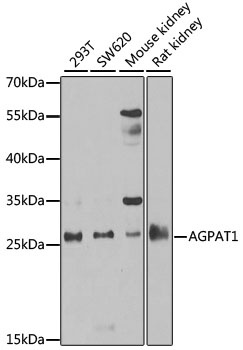
Western blot analysis of various lysates using AGPAT1 Rabbit pAb (CAB6517) at 1:1000 dilution. Secondary antibody: HRP-conjugated Goat anti-Rabbit IgG (H+L) (AS014) at 1:10000 dilution. Lysates/proteins: 25μg per lane. Blocking buffer: 3% nonfat dry milk in TBST. Detection: ECL Basic Kit (AbGn00020). Exposure time: 90s.
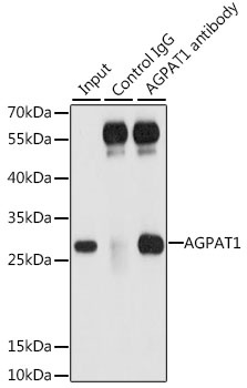
Immunoprecipitation analysis of 200 μg extracts of 293T cells, using 3 μg AGPAT1 antibody (CAB6517). Western blot was performed from the immunoprecipitate using AGPAT1 antibody (CAB6517) at a dilution of 1:1000.