The AGR2 Antibody (CAB7064) is a high-quality antibody developed for reliable detection and analysis of target proteins. This antibody, produced in rabbits, exhibits high specificity and sensitivity for human samples, making it ideal for use in Western blot applications. By targeting the AGR2 protein, this antibody enables precise detection and analysis in a variety of cell types, providing insights into the role of AGR2 in cancer biology.AGR2, a member of the protein disulfide isomerase family, is overexpressed in many types of cancer and is believed to promote tumor growth, invasion, and resistance to therapy.
This antibody is validated for use in WB, IHC-P, IF/ICC, ELISA applications and has demonstrated reactivity against Human, Mouse, Rat samples.
Product Name:
AGR2 Antibody
SKU:
CAB7064
Size:
20μL, 100μL
Reactivity:
Human, Mouse, Rat
Conjugate:
Unconjugated
Immunogen:
Recombinant protein (or fragment).This information is considered to be commercially sensitive.
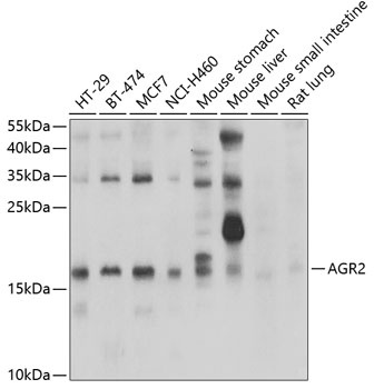
HT-29, BT-474, MCF7, NCI-H460, Mouse stomach, Mouse liver, Mouse small intestine, Rat lung
Cellular Localization:
Endoplasmic Reticulum, Secreted.
Calculated MW:
20kDa
Observed MW:
20kDa
This gene encodes a member of the disulfide isomerase (PDI) family of endoplasmic reticulum (ER) proteins that catalyze protein folding and thiol-disulfide interchange reactions. The encoded protein has an N-terminal ER-signal sequence, a catalytically active thioredoxin domain, and a C-terminal ER-retention sequence. This protein plays a role in cell migration, cellular transformation and metastasis and is as a p53 inhibitor. As an ER-localized molecular chaperone, it plays a role in the folding, trafficking, and assembly of cysteine-rich transmembrane receptors and the cysteine-rich intestinal gylcoprotein mucin. This gene has been implicated in inflammatory bowel disease and cancer progression.
Purification Method
Affinity purification
Gene ID
10551
RRID
AB_2767619
Buffer Information
Store at -20℃. Avoid freeze / thaw cycles. Buffer: PBS with 0.09% Sodium azide,50% glycerol,pH7.3.
Western blot analysis of various lysates using AGR2 Rabbit pAb (CAB7064) at 1:1000 dilution. Secondary antibody: HRP-conjugated Goat anti-Rabbit IgG (H+L) (CABS014) at 1:10000 dilution. Lysates/proteins: 25μg per lane. Blocking buffer: 3% nonfat dry milk in TBST. Detection: ECL Basic Kit (AbGn00020). Exposure time: 1s.
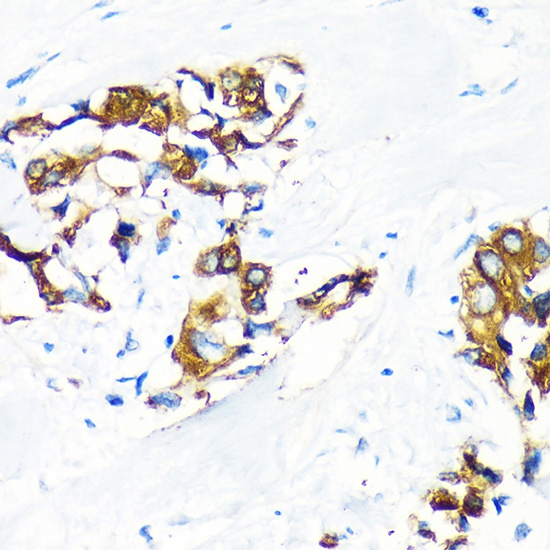
Immunohistochemistry analysis of paraffin-embedded Human lung cancer using AGR2 Rabbit pAb (CAB7064) at dilution of 1:100 (40x lens). Microwave antigen retrieval performed with 0.01M PBS Buffer (pH 7.2) prior to IHC staining.
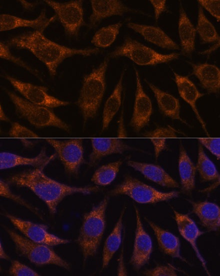
Immunofluorescence analysis of L929 cells using AGR2 Rabbit pAb (CAB7064) at dilution of 1:100. Secondary antibody: Cy3-conjugated Goat anti-Rabbit IgG (H+L) (CABS007) at 1:500 dilution. Blue: DAPI for nuclear staining.
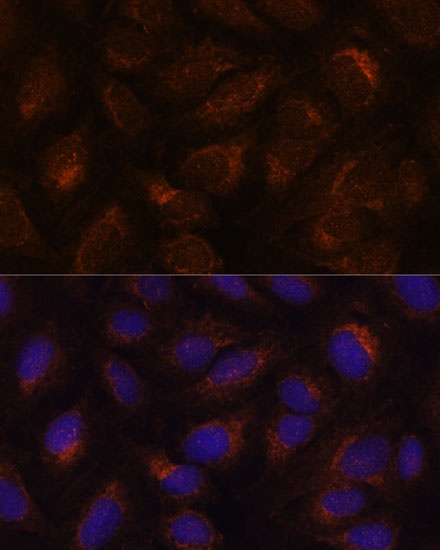
Immunofluorescence analysis of U-2 OS cells using AGR2 Rabbit pAb (CAB7064) at dilution of 1:100. Secondary antibody: Cy3-conjugated Goat anti-Rabbit IgG (H+L) (CABS007) at 1:500 dilution. Blue: DAPI for nuclear staining.