The Iba1 Antibody (CAB12391) is a high-quality antibody developed for reliable detection and analysis of target proteins. This gene encodes a protein that binds actin and calcium. This gene is induced by cytokines and interferon and may promote macrophage activation and growth of vascular smooth muscle cells and T-lymphocytes. Polymorphisms in this gene may be associated with systemic sclerosis. Alternative splicing results in multiple transcript variants, but the full-length and coding nature of some of these variants is not certain. RRID AB_2759236 Gene ID 199 Swiss Prot Synonym IBA1; IRT1; AIF-1; IRT-1; AIF1/IBA1
This antibody is validated for use in WB, ELISA, IF-P applications and has demonstrated reactivity against Human, Mouse, Rat samples.
Product Name:
Iba1 Antibody
SKU:
CAB12391
Size:
100μL, 20μL
Reactivity:
Human, Mouse, Rat
Clone Number:
-
Conjugate:
Unconjugated
Immunogen:
Recombinant protein (or fragment).This information is considered to be commercially sensitive.
Tested Applications:
WBELISAIF-P
Recommended Dilution:
WB
1:1000 - 1:5000
IF-P
1:50 - 1:200
ELISA
Recommended starting concentration is 1 μg/mL. Please optimize the concentration based on your specific assay requirements.
This gene encodes a protein that binds actin and calcium. This gene is induced by cytokines and interferon and may promote macrophage activation and growth of vascular smooth muscle cells and T-lymphocytes. Polymorphisms in this gene may be associated with systemic sclerosis. Alternative splicing results in multiple transcript variants, but the full-length and coding nature of some of these variants is not certain. RRID AB_2759236 Gene ID 199 Swiss Prot Synonym IBA1; IRT1; AIF-1; IRT-1; AIF1/IBA1
Purification Method:
Affinity purification
Gene ID:
199
RRID:
AB_2759236
Buffer Information:
Store at -20℃. Avoid freeze / thaw cycles. Buffer: PBS containing 50% glycerol, preserved with proclin300 or sodium azide, pH 7.3.
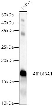
Western blot analysis of lysates from THP-1 cells, using AIF1/IBA1 Rabbit pAb (CAB12391) at 1:2000 dilution. Secondary antibody: HRP-conjugated Goat anti-Rabbit IgG (H+L) (AS014) at 1:10000 dilution. Lysates/proteins: 25μg per lane. Blocking buffer: 3% nonfat dry milk in TBST. Detection: ECL Basic Kit (AbGn00020). Exposure time: 30s.
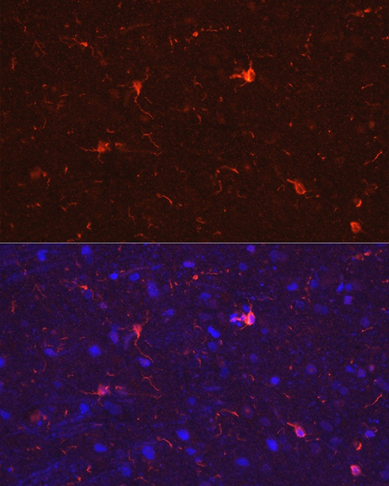
Immunofluorescence analysis of paraffin-embedded rat brain using AIF1/IBA1 Rabbit pAb (CAB12391) at dilution of 1:50 (40x lens). Secondary antibody: Cy3-conjugated Goat anti-Rabbit IgG (H+L) (AS007) at 1:500 dilution. Blue: DAPI for nuclear staining.