The AIF Polyclonal Antibody (CAB0811) is a high-quality antibody developed for reliable detection and analysis of target proteins. This antibody, produced in rabbits, is highly reactive with human samples and has been validated for use in various applications such as Western blot and immunofluorescence.AIF, or apoptosis-inducing factor, is a mitochondrial protein that plays a crucial role in the regulation of programmed cell death. Dysregulation of AIF has been implicated in various diseases, including neurodegenerative disorders and cancers. By using this antibody to detect and analyze AIF expression in different cell types, researchers can gain valuable insights into the molecular mechanisms underlying these diseases.
This antibody is validated for use in WB, IHC-P, IF/ICC, ELISA, IF-P applications and has demonstrated reactivity against Human, Mouse, Rat samples.
Product Name:
AIF Polyclonal Antibody
SKU:
CAB0811
Size:
20μL, 100μL
Reactivity:
Human, Mouse, Rat
Conjugate:
Unconjugated
Immunogen:
Recombinant protein (or fragment).This information is considered to be commercially sensitive.
This gene encodes a flavoprotein essential for nuclear disassembly in apoptotic cells, and it is found in the mitochondrial intermembrane space in healthy cells. Induction of apoptosis results in the translocation of this protein to the nucleus where it affects chromosome condensation and fragmentation. In addition, this gene product induces mitochondria to release the apoptogenic proteins cytochrome c and caspase-9. Mutations in this gene cause combined oxidative phosphorylation deficiency 6 (COXPD6), a severe mitochondrial encephalomyopathy, as well as Cowchock syndrome, also known as X-linked recessive Charcot-Marie-Tooth disease-4 (CMTX-4), a disorder resulting in neuropathy, and axonal and motor-sensory defects with deafness and cognitive disability. Alternative splicing results in multiple transcript variants. A related pseudogene has been identified on chromosome 10.
Purification Method
Affinity purification
Gene ID
9131
RRID
AB_2757407
Buffer Information
Store at -20℃. Avoid freeze / thaw cycles. Buffer: PBS containing 50% glycerol, preserved with proclin300 or sodium azide, pH 7.3.
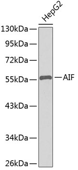
Western blot analysis of lysates from HepG2 cells, using AIF Rabbit pAb (CAB0811). Secondary antibody: HRP-conjugated Goat anti-Rabbit IgG (H+L) (CABS014) at 1:10000 dilution. Lysates/proteins: 25μg per lane. Blocking buffer: 3% nonfat dry milk in TBST.
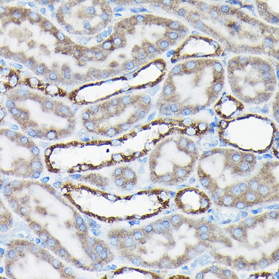
Immunohistochemistry analysis of paraffin-embedded Rat kidney using AIF Rabbit pAb (CAB0811) at dilution of 1:50 (40x lens). High pressure antigen retrieval performed with 0.01M Citrate buffer (pH 6.0) prior to IHC staining.
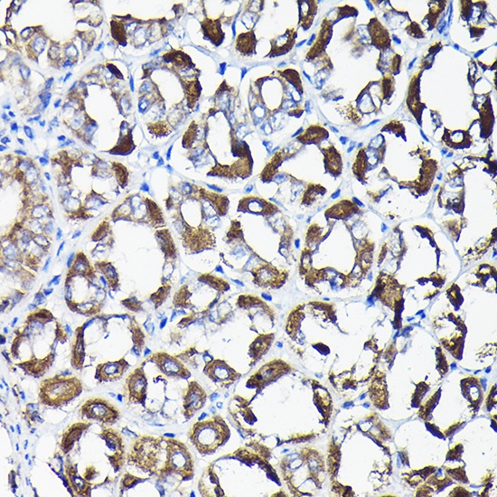
Immunohistochemistry analysis of paraffin-embedded Mouse stomach using AIF Rabbit pAb (CAB0811) at dilution of 1:50 (40x lens). High pressure antigen retrieval performed with 0.01M Citrate buffer (pH 6.0) prior to IHC staining.
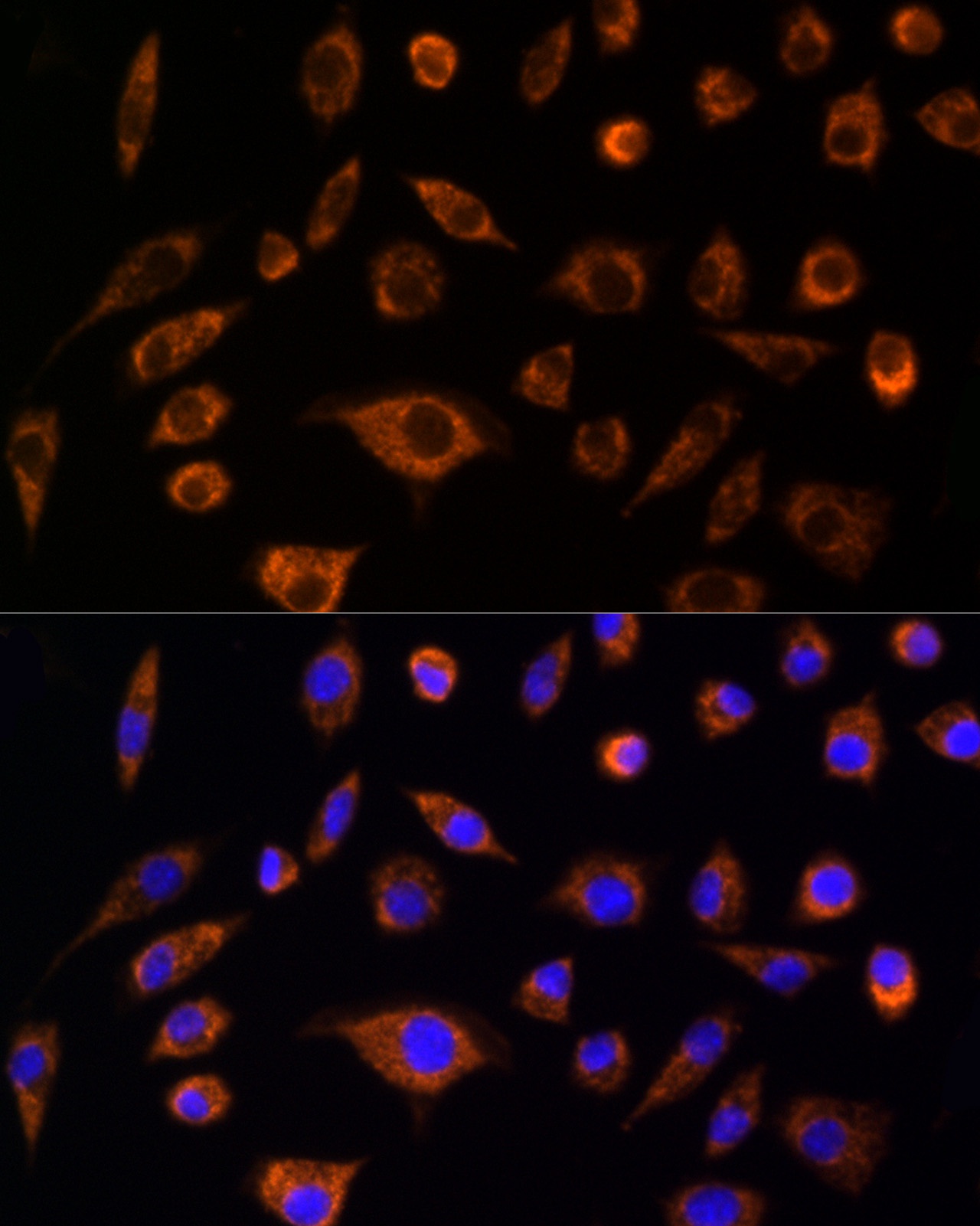
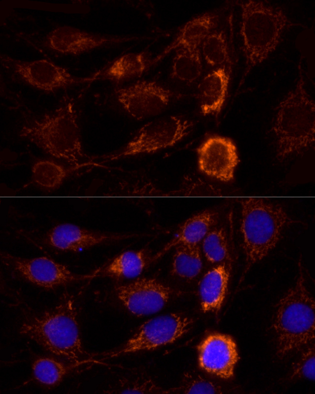
Immunofluorescence analysis of L929 cells using AIF Rabbit pAb (CAB0811) at dilution of 1:100 (40x lens). Secondary antibody: Cy3-conjugated Goat anti-Rabbit IgG (H+L) (CABS007) at 1:500 dilution. Blue: DAPI for nuclear staining.
Immunofluorescence analysis of C6 cells using AIF Rabbit pAb (CAB0811) at dilution of 1:100 (40x lens). Secondary antibody: Cy3-conjugated Goat anti-Rabbit IgG (H+L) (CABS007) at 1:500 dilution. Blue: DAPI for nuclear staining.
. Secondary antibody: HRP Goat Anti-Rabbit IgG (H+L) at 1:10000 dilution. Lysates/proteins: 25μg per lane. Blocking buffer: 3% nonfat dry milk in TBST.")
. Secondary antibody: HRP Goat Anti-Rabbit IgG (H+L) at 1:10000 dilution. Lysates/proteins: 25μg per lane. Blocking buffer: 3% nonfat dry milk in TBST.")


