The AIM2 Antibody (CAB3356) is a high-quality antibody developed for reliable detection and analysis of target proteins. This antibody, produced through immunization of rabbits, exhibits high reactivity with human samples, making it an ideal choice for use in Western blot applications. By specifically binding to the AIM2 protein, this antibody enables precise detection and analysis in various cell types, allowing for in-depth investigations in the fields of immunology and cancer research.AIM2, also known as absent in melanoma 2, plays a crucial role in immune surveillance and inflammatory responses by detecting cytoplasmic DNA and initiating the inflammasome pathway.
This antibody is validated for use in WB, IF/ICC, ELISA applications and has demonstrated reactivity against Human, Mouse, Rat samples.
Product Name:
AIM2 Antibody
SKU:
CAB3356
Size:
20μL, 100μL
Reactivity:
Human, Mouse, Rat
Conjugate:
Unconjugated
Immunogen:
Synthetic peptide. This information is considered to be commercially sensitive.
Recommended starting concentration is 1 μg/mL. Please optimize the concentration based on your specific assay requirements.
Synonyms:
PYHIN4, AIM2
Positive Sample:
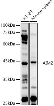
HT-29, Mouse spleen
Cellular Localization:
Cytoplasm, Nucleus.
Calculated MW:
39kDa
Observed MW:
39kDa
AIM2 is a member of the IFI20X /IFI16 family. It plays a putative role in tumorigenic reversion and may control cell proliferation. Interferon-gamma induces expression of AIM2.
Purification Method
Affinity purification
Gene ID
9447
RRID
AB_2765071
Buffer Information
Store at -20℃. Avoid freeze / thaw cycles. Buffer: PBS containing 50% glycerol, preserved with proclin300 or sodium azide, pH 7.3.
Western blot analysis of various lysates using AIM2 Rabbit pAb (CAB3356) at 1:1000 dilution. Secondary antibody: HRP-conjugated Goat anti-Rabbit IgG (H+L) (CABS014) at 1:10000 dilution. Lysates/proteins: 25μg per lane. Blocking buffer: 3% nonfat dry milk in TBST. Detection: ECL Basic Kit (AbGn00020). Exposure time: 30s.
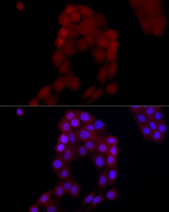
Immunofluorescence analysis of A-431 cells using AIM2 Rabbit pAb (CAB3356) at dilution of 1:50 (40x lens). Secondary antibody: Cy3-conjugated Goat anti-Rabbit IgG (H+L) (CABS007) at 1:500 dilution. Blue: DAPI for nuclear staining.