The AIPL1 Antibody (CAB6458) is a high-quality antibody developed for reliable detection and analysis of target proteins. This antibody, produced in rabbits, is highly specific for human samples and is validated for Western blot applications.AIPL1 is a key protein involved in the maintenance and function of photoreceptor cells in the retina, making it essential for normal vision. Mutations in the AIPL1 gene are known to cause LCA, highlighting the importance of studying this protein for understanding the mechanisms underlying this vision impairment disorder.
This antibody is validated for use in WB, IF/ICC, ELISA applications and has demonstrated reactivity against Human, Mouse samples.
Product Name:
AIPL1 Antibody
SKU:
CAB6458
Size:
20μL, 100μL
Reactivity:
Human, Mouse
Conjugate:
Unconjugated
Immunogen:
Recombinant protein (or fragment).This information is considered to be commercially sensitive.
Recommended starting concentration is 1 μg/mL. Please optimize the concentration based on your specific assay requirements.
Synonyms:
LCA4, AIPL2, AIPL1
Positive Sample:
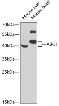
Mouse liver, Mouse heart
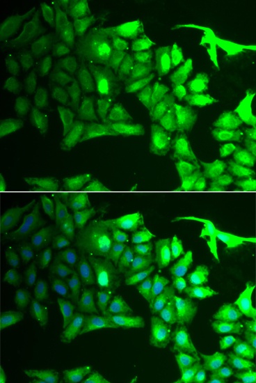
Cellular Localization:
Cytoplasm, Nucleus.
Calculated MW:
44kDa
Observed MW:
40-45kDa
Leber congenital amaurosis (LCA) is the most severe inherited retinopathy with the earliest age of onset and accounts for at least 5% of all inherited retinal diseases. Affected individuals are diagnosed at birth or in the first few months of life with nystagmus, severely impaired vision or blindness and an abnormal or flat electroretinogram. The photoreceptor/pineal-expressed gene, AIPL1, encoding aryl-hydrocarbon interacting protein-like 1, is located within the LCA4 candidate region. The encoded protein contains three tetratricopeptide motifs, consistent with chaperone or nuclear transport activity. Mutations in this gene may cause approximately 20% of recessive LCA. Alternative splicing results in multiple transcript variants.
Purification Method
Affinity purification
Gene ID
23746
RRID
AB_2767060
Buffer Information
Store at -20℃. Avoid freeze / thaw cycles. Buffer: PBS containing 50% glycerol, preserved with proclin300 or sodium azide, pH 7.3.
Western blot analysis of various lysates using AIPL1 Rabbit pAb (CAB6458) at 1:1000 dilution. Secondary antibody: HRP-conjugated Goat anti-Rabbit IgG (H+L) (CABS014) at 1:10000 dilution. Lysates/proteins: 25μg per lane. Blocking buffer: 3% nonfat dry milk in TBST. Detection: ECL Basic Kit (AbGn00020). Exposure time: 90s.
Immunofluorescence analysis of HeLa cells using AIPL1 Rabbit pAb (CAB6458). Secondary antibody: Cy3-conjugated Goat anti-Rabbit IgG (H+L) (CABS007) at 1:500 dilution. Blue: DAPI for nuclear staining.