The ALDH1L1 Antibody (CAB7067) is a high-quality antibody developed for reliable detection and analysis of target proteins. This antibody, raised in rabbits, exhibits high reactivity with human samples and has been validated for use in Western blot applications. By binding specifically to the ALDH1L1 protein, this antibody enables accurate detection and analysis in various cell types, making it ideal for studies in cancer biology and neuroscience.ALDH1L1, also known as mitochondrial 10-formyltetrahydrofolate dehydrogenase, plays a crucial role in maintaining cellular folate levels and nucleotide synthesis, making it a potential target for cancer therapy.
This antibody is validated for use in WB, ELISA applications and has demonstrated reactivity against Human, Mouse, Rat samples.
Product Name:
ALDH1L1 Antibody
SKU:
CAB7067
Size:
20μL, 100μL
Reactivity:
Human, Mouse, Rat
Conjugate:
Unconjugated
Immunogen:
Recombinant protein (or fragment).This information is considered to be commercially sensitive.
Recommended starting concentration is 1 μg/mL. Please optimize the concentration based on your specific assay requirements.
Synonyms:
FDH, FTHFD, 10-fTHF, 10-FTHFDH, ALDH1L1
Positive Sample:
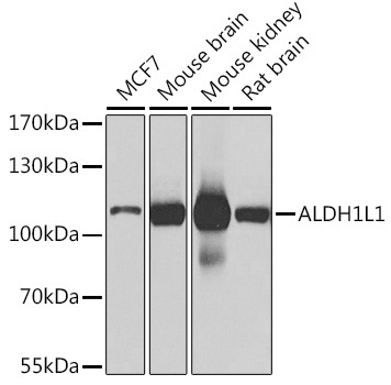
MCF7, Mouse brain, Mouse kidney, Rat brain
Cellular Localization:
Cytoplasm.
Calculated MW:
99kDa
Observed MW:
120kDa
The protein encoded by this gene catalyzes the conversion of 10-formyltetrahydrofolate, nicotinamide adenine dinucleotide phosphate (NADP+), and water to tetrahydrofolate, NADPH, and carbon dioxide. The encoded protein belongs to the aldehyde dehydrogenase family. Loss of function or expression of this gene is associated with decreased apoptosis, increased cell motility, and cancer progression. There is an antisense transcript that overlaps on the opposite strand with this gene locus. Alternative splicing results in multiple transcript variants.
Purification Method
Affinity purification
Gene ID
10840
RRID
AB_2767622
Buffer Information
Store at -20℃. Avoid freeze / thaw cycles. Buffer: PBS containing 50% glycerol, preserved with proclin300 or sodium azide, pH 7.3.
Western blot analysis of various lysates using ALDH1L1 Rabbit pAb (CAB7067) at 1:1000 dilution. Secondary antibody: HRP-conjugated Goat anti-Rabbit IgG (H+L) (CABS014) at 1:10000 dilution. Lysates/proteins: 25μg per lane. Blocking buffer: 3% nonfat dry milk in TBST. Detection: ECL Basic Kit (AbGn00020). Exposure time: 1s.