The ALDH7A1 Antibody (CAB8629) is a high-quality antibody developed for reliable detection and analysis of target proteins. This antibody, produced in rabbits, is highly specific for human samples and is validated for use in Western blot applications. By targeting the ALDH7A1 protein, this antibody allows for the detection and analysis of ALDH7A1 expression in a variety of cell types, making it suitable for studies in metabolism, neurology, and genetic disorders.ALDH7A1, also known as antiquitin, plays a crucial role in the detoxification of harmful aldehydes and the biosynthesis of amino acids such as lysine and tryptophan.
This antibody is validated for use in WB, IHC-P, IF/ICC, ELISA applications and has demonstrated reactivity against Human, Mouse, Rat samples.
Product Name:
ALDH7A1 Antibody
SKU:
CAB8629
Size:
20μL, 100μL
Reactivity:
Human, Mouse, Rat
Conjugate:
Unconjugated
Immunogen:
Recombinant protein (or fragment).This information is considered to be commercially sensitive.
Recommended starting concentration is 1 μg/mL. Please optimize the concentration based on your specific assay requirements.
Synonyms:
EPD, PDE, ATQ1, ALDH7A1
Positive Sample:
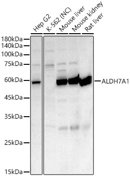
Hep G2, Mouse liver, Mouse kidney, Rat liver
Cellular Localization:
Cytoplasm, Mitochondrion, Nucleus, Cytosol.
Calculated MW:
58kDa
Observed MW:
58kDa
The protein encoded by this gene is a member of subfamily 7 in the aldehyde dehydrogenase gene family. These enzymes are thought to play a major role in the detoxification of aldehydes generated by alcohol metabolism and lipid peroxidation. This particular member has homology to a previously described protein from the green garden pea, the 26g pea turgor protein. It is also involved in lysine catabolism that is known to occur in the mitochondrial matrix. Recent reports show that this protein is found both in the cytosol and the mitochondria, and the two forms likely arise from the use of alternative translation initiation sites. An additional variant encoding a different isoform has also been found for this gene. Mutations in this gene are associated with pyridoxine-dependent epilepsy. Several related pseudogenes have also been identified.
Purification Method
Affinity purification
Gene ID
501
RRID
AB_2768312
Buffer Information
Store at -20℃. Avoid freeze / thaw cycles. Buffer: PBS containing 50% glycerol, preserved with proclin300 or sodium azide, pH 7.3.
Western blot analysis of various lysates, using ALDH7A1 Rabbit pAb (CAB8629) at 1:400 dilution. Secondary antibody: HRP-conjugated Goat anti-Rabbit IgG (H+L) (CABS014) at 1:10000 dilution. Lysates/proteins: 25μg per lane. Blocking buffer: 3% nonfat dry milk in TBST. Detection: ECL Enhanced Kit (AbGn00021). Negative control (NC): K-562. Exposure time: 75s.
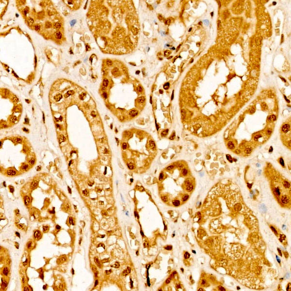
Immunohistochemistry analysis of paraffin-embedded Human kidney tissue using ALDH7A1 Rabbit pAb (CAB8629) at a dilution of 1:50 (40x lens). High pressure antigen retrieval performed with 0.01M Citrate buffer (pH 6.0) prior to IHC staining.
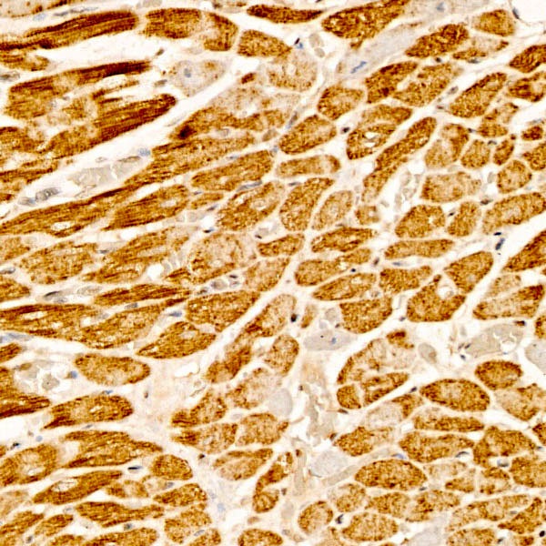
Immunohistochemistry analysis of paraffin-embedded Rat heart tissue using ALDH7A1 Rabbit pAb (CAB8629) at a dilution of 1:50 (40x lens). High pressure antigen retrieval performed with 0.01M Citrate buffer (pH 6.0) prior to IHC staining.
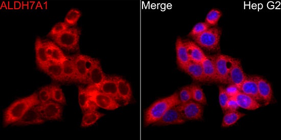
Immunofluorescence analysis of HepG2 cells using ALDH7A1 Rabbit pAb (CAB8629) at dilution of 1:50 (40x lens). Secondary antibody: Cy3-conjugated Goat anti-Rabbit IgG (H+L) (CABS007) at 1:500 dilution. Blue: DAPI for nuclear staining.


 at 1:10000 dilution. Lysates/proteins: 25ug per lane. Blocking buffer: 3% nonfat dry milk in TBST. Detection: ECL Basic Kit. Exposure time: 10s.")


