The AMPD3 Antibody (CAB6354) is a high-quality antibody developed for reliable detection and analysis of target proteins. This antibody, produced in rabbits, shows high reactivity with human samples and is validated for use in Western blot applications. By specifically targeting AMP Deaminase 3, researchers can efficiently detect and analyze this protein in a variety of cell types, making it an excellent choice for studies in biochemistry and molecular biology.
This antibody is validated for use in WB, ELISA applications and has demonstrated reactivity against Human, Mouse, Rat samples.
Product Name:
AMPD3 Antibody
SKU:
CAB6354
Size:
20μL, 100μL
Reactivity:
Human, Mouse, Rat
Conjugate:
Unconjugated
Immunogen:
Recombinant protein (or fragment).This information is considered to be commercially sensitive.
Recommended starting concentration is 1 μg/mL. Please optimize the concentration based on your specific assay requirements.
Synonyms:
AMPD3
Positive Sample:
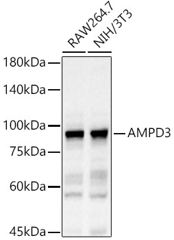
RAW264.7, NIH/3T3
Cellular Localization:
Cytosol, Extracellular Region.
Calculated MW:
89kDa
Observed MW:
89kDa
This gene encodes a member of the AMP deaminase gene family. The encoded protein is a highly regulated enzyme that catalyzes the hydrolytic deamination of adenosine monophosphate to inosine monophosphate, a branch point in the adenylate catabolic pathway. This gene encodes the erythrocyte (E) isoforms, whereas other family members encode isoforms that predominate in muscle (M) and liver (L) cells. Mutations in this gene lead to the clinically asymptomatic, autosomal recessive condition erythrocyte AMP deaminase deficiency. Alternatively spliced transcript variants encoding different isoforms of this gene have been described.
Purification Method
Affinity purification
Gene ID
272
RRID
AB_2766956
Buffer Information
Store at -20℃. Avoid freeze / thaw cycles. Buffer: PBS containing 50% glycerol, preserved with proclin300 or sodium azide, pH 7.3.
Western blot analysis of various lysates, using AMPD3 Rabbit pAb (CAB6354) at 1:1500 dilution. Secondary antibody: HRP-conjugated Goat anti-Rabbit IgG (H+L) (CABS014) at 1:10000 dilution. Lysates/proteins: 25μg per lane. Blocking buffer: 3% nonfat dry milk in TBST. Detection: ECL Basic Kit (AbGn00020). Exposure time: 60s.