The AMPH Antibody (CAB5389) is a high-quality antibody developed for reliable detection and analysis of target proteins. This antibody, produced in rabbits, is highly specific to human samples and has been validated for use in immunohistochemistry and Western blotting applications.Amphiphysin is known to play a key role in synaptic vesicle endocytosis, making it important for studying neurobiology and neurodegenerative diseases. By binding specifically to the amphiphysin protein, this antibody enables researchers to detect and analyze its expression in various cell types and tissues.
This antibody is validated for use in WB, IF/ICC, ELISA applications and has demonstrated reactivity against Human, Mouse, Rat samples.
Product Name:
AMPH Antibody
SKU:
CAB5389
Size:
20μL, 100μL
Reactivity:
Human, Mouse, Rat
Conjugate:
Unconjugated
Immunogen:
Recombinant protein (or fragment).This information is considered to be commercially sensitive.
This gene encodes a protein associated with the cytoplasmic surface of synaptic vesicles. A subset of patients with stiff-man syndrome who were also affected by breast cancer are positive for autoantibodies against this protein. Alternate splicing of this gene results in two transcript variants encoding different isoforms. Additional splice variants have been described, but their full length sequences have not been determined. A pseudogene of this gene is found on chromosome 11.
Purification Method
Affinity purification
Gene ID
273
RRID
AB_2766198
Buffer Information
Store at -20℃. Avoid freeze / thaw cycles. Buffer: PBS with 0.01% thimerosal,50% glycerol,pH7.3.
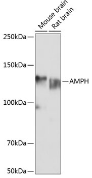
Western blot analysis of various lysates using AMPH Rabbit pAb (CAB5389) at 1:1000 dilution. Secondary antibody: HRP-conjugated Goat anti-Rabbit IgG (H+L) (CABS014) at 1:10000 dilution. Lysates/proteins: 25μg per lane. Blocking buffer: 3% nonfat dry milk in TBST. Detection: ECL Basic Kit (AbGn00020). Exposure time: 1s.
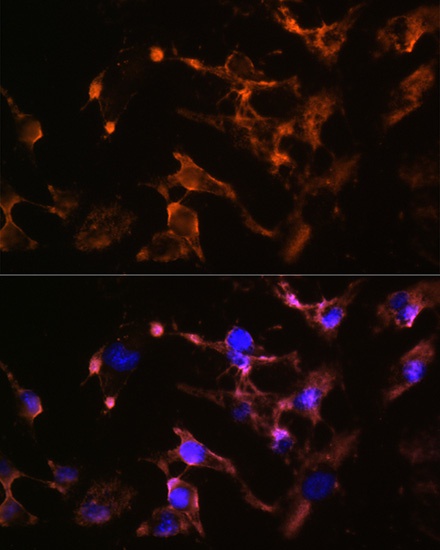
Immunofluorescence analysis of U-251 MG cells using AMPH Rabbit pAb (CAB5389) at dilution of 1:100 (40x lens). Secondary antibody: Cy3-conjugated Goat anti-Rabbit IgG (H+L) (CABS007) at 1:500 dilution. Blue: DAPI for nuclear staining.