The ANGPTL4 Antibody (CAB2011) is a high-quality antibody developed for reliable detection and analysis of target proteins. This antibody, produced in rabbits, exhibits high reactivity with human samples and is validated for use in Western blot applications. By binding to the ANGPTL4 protein, this antibody enables accurate detection and analysis in various cell types, making it an excellent tool for studies in metabolic disorders, cardiovascular diseases, and cancer research.
This antibody is validated for use in WB, IHC-P, IF/ICC, ELISA applications and has demonstrated reactivity against Human, Mouse, Rat samples.
Product Name:
ANGPTL4 Antibody
SKU:
CAB2011
Size:
20μL, 100μL
Reactivity:
Human, Mouse, Rat
Conjugate:
Unconjugated
Immunogen:
Recombinant protein (or fragment).This information is considered to be commercially sensitive.
This gene encodes a glycosylated, secreted protein containing a C-terminal fibrinogen domain. The encoded protein is induced by peroxisome proliferation activators and functions as a serum hormone that regulates glucose homeostasis, lipid metabolism, and insulin sensitivity. This protein can also act as an apoptosis survival factor for vascular endothelial cells and can prevent metastasis by inhibiting vascular growth and tumor cell invasion. The C-terminal domain may be proteolytically-cleaved from the full-length secreted protein. Decreased expression of this gene has been associated with type 2 diabetes. Alternative splicing results in multiple transcript variants. This gene was previously referred to as ANGPTL2 but has been renamed ANGPTL4.
Purification Method
Affinity purification
Gene ID
51129
RRID
AB_2764035
Buffer Information
Store at -20℃. Avoid freeze / thaw cycles. Buffer: PBS containing 50% glycerol, preserved with proclin300 or sodium azide, pH 7.3.
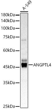
Western blot analysis of lysates from A-549 cells, using ANGPTL4 Rabbit pAb (CAB2011) at 1:600 dilution. Secondary antibody: HRP-conjugated Goat anti-Rabbit IgG (H+L) (CABS014) at 1:10000 dilution. Lysates/proteins: 25μg per lane. Blocking buffer: 3% nonfat dry milk in TBST. Detection: ECL Basic Kit (AbGn00020). Exposure time: 5s.
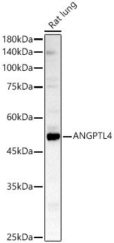
Western blot analysis of lysates from Rat lung, using ANGPTL4 Rabbit pAb (CAB2011) at 1:600 dilution. Secondary antibody: HRP-conjugated Goat anti-Rabbit IgG (H+L) (CABS014) at 1:10000 dilution. Lysates/proteins: 25μg per lane. Blocking buffer: 3% nonfat dry milk in TBST. Detection: ECL Basic Kit (AbGn00020). Exposure time: 180s.
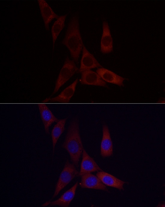
Immunofluorescence analysis of NIH/3T3 cells using ANGPTL4 Rabbit pAb (CAB2011) at dilution of 1:50 (40x lens). Secondary antibody: Cy3-conjugated Goat anti-Rabbit IgG (H+L) (CABS007) at 1:500 dilution. Blue: DAPI for nuclear staining.