The 67kDa Laminin Receptor Monoclonal Antibody (CAB5968) is a high-quality antibody developed for reliable detection and analysis of target proteins. This polyclonal antibody, produced in rabbits, is highly specific and reacts strongly with human samples. It is suitable for use in techniques such as Western blotting, immunohistochemistry, and immunofluorescence.The 67kDa laminin receptor, also known as LRP/LR or RPSA, is a multifunctional protein involved in various cellular processes, including cell adhesion, migration, and signaling. It has been implicated in cancer progression, metastasis, and angiogenesis, making it a promising target for cancer research and therapeutics.
This antibody is validated for use in WB, IF/ICC, ELISA applications and has demonstrated reactivity against Human, Mouse, Rat samples.
Product Name:
67kDa Laminin Receptor Monoclonal Antibody
SKU:
CAB5968
Size:
20μL, 100μL
Reactivity:
Human, Mouse, Rat
Clone Number:
ARC2109
Conjugate:
Unconjugated
Immunogen:
Synthetic peptide. This information is considered to be commercially sensitive.
Laminins, a family of extracellular matrix glycoproteins, are the major noncollagenous constituent of basement membranes. They have been implicated in a wide variety of biological processes including cell adhesion, differentiation, migration, signaling, neurite outgrowth and metastasis. Many of the effects of laminin are mediated through interactions with cell surface receptors. These receptors include members of the integrin family, as well as non-integrin laminin-binding proteins. This gene encodes a high-affinity, non-integrin family, laminin receptor 1. This receptor has been variously called 67 kD laminin receptor, 37 kD laminin receptor precursor (37LRP) and p40 ribosome-associated protein. The amino acid sequence of laminin receptor 1 is highly conserved through evolution, suggesting a key biological function. It has been observed that the level of the laminin receptor transcript is higher in colon carcinoma tissue and lung cancer cell line than their normal counterparts. Also, there is a correlation between the upregulation of this polypeptide in cancer cells and their invasive and metastatic phenotype. Multiple copies of this gene exist, however, most of them are pseudogenes thought to have arisen from retropositional events. Two alternatively spliced transcript variants encoding the same protein have been found for this gene.
Purification Method
Affinity purification
Gene ID
3921
Buffer Information
Store at -20℃. Avoid freeze / thaw cycles. Buffer: PBS containing 50% glycerol and 0.05% BSA, preserved with proclin300 or sodium azide, pH 7.3.
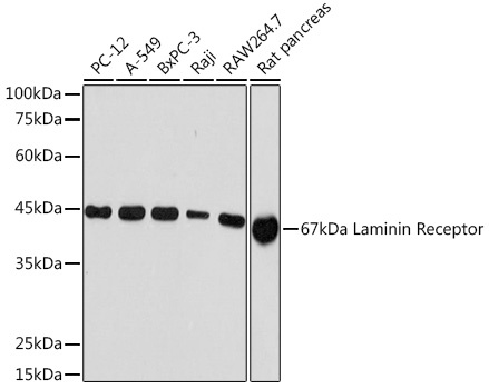
Western blot analysis of various lysates using 67kDa Laminin Receptor Rabbit mAb (CAB5968) at 1:1000 dilution. Secondary antibody: HRP-conjugated Goat anti-Rabbit IgG (H+L) (CABS014) at 1:10000 dilution. Lysates/proteins: 25μg per lane. Blocking buffer: 3% nonfat dry milk in TBST. Detection: ECL Basic Kit (AbGn00020). Exposure time: 10s.
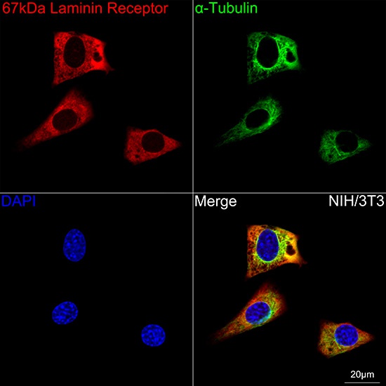
Confocal imaging of NIH/3T3 cells using 67kDa Laminin Receptor Rabbit mAb (CAB5968, dilution 1:100) followed by a further incubation with Cy3 Goat Anti-Rabbit IgG (H+L) (CABS007, dilution 1:500) (Red). The cells were counterstained with α-Tubulin Mouse mAb (AC012, dilution 1:400) followed by incubation with ABflo® 488-conjugated Goat Anti-Mouse IgG (H+L) Ab (CABS076, dilution 1:500) (Green). DAPI was used for nuclear staining (Blue). Objective: 100x.