The A-RAF Monoclonal Antibody (CAB8687) is a high-quality antibody developed for reliable detection and analysis of target proteins. Enables protein serine/threonine kinase activity. Involved in negative regulation of apoptotic process; regulation of TOR signaling; and regulation of cellular protein metabolic process. Predicted to be active in cytosol and mitochondrion. Biomarker of high grade glioma.
This antibody is validated for use in WB, IHC-P, ELISA applications and has demonstrated reactivity against Human samples.
Product Name:
A-RAF Monoclonal Antibody
SKU:
CAB8687
Size:
100μL, 20μL
Reactivity:
Human
Clone Number:
ARC1782
Conjugate:
Unconjugated
Immunogen:
Recombinant protein (or fragment).This information is considered to be commercially sensitive.
Tested Applications:
WBIHC-PELISA
Recommended Dilution:
WB
1:500 - 1:1000
IHC-P
1:50 - 1:200
ELISA
Recommended starting concentration is 1 μg/mL. Please optimize the concentration based on your specific assay requirements.
Synonyms:
PKS2, A-RAF, ARAF1, RAFA1
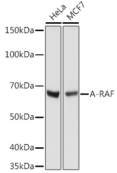
Positive Sample:
HeLa, MCF7
Cellular Localization:
Cytosol, Mitochondrion.
Calculated MW:
68kDa
Observed MW:
68kDa
Enables protein serine/threonine kinase activity. Involved in negative regulation of apoptotic process; regulation of TOR signaling; and regulation of cellular protein metabolic process. Predicted to be active in cytosol and mitochondrion. Biomarker of high grade glioma.
Purification Method
Affinity purification
Gene ID
369
Buffer Information
Store at -20℃. Avoid freeze / thaw cycles. Buffer: PBS containing 50% glycerol and 0.05% BSA, preserved with proclin300 or sodium azide, pH 7.3.
Western blot analysis of various lysates using A-RAF Rabbit mAb (CAB8687) at 1:1000 dilution. Secondary antibody: HRP-conjugated Goat anti-Rabbit IgG (H+L) (AS014) at 1:10000 dilution. Lysates/proteins: 25μg per lane. Blocking buffer: 3% nonfat dry milk in TBST. Detection: ECL Basic Kit (AbGn00020). Exposure time: 10s.
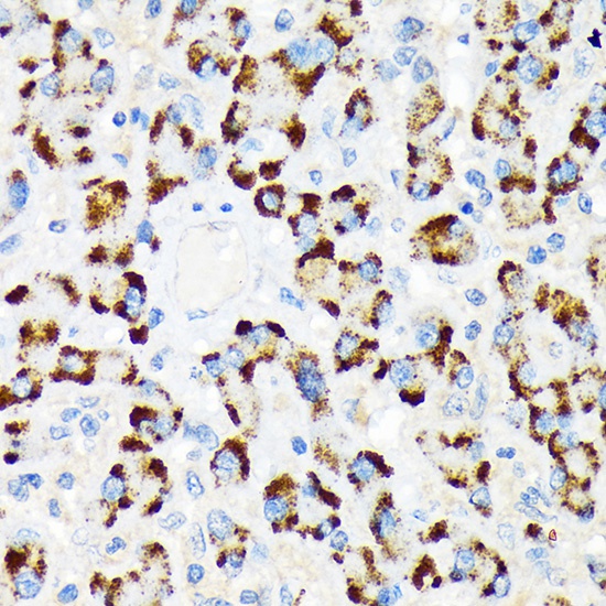
Immunohistochemistry analysis of paraffin-embedded Human liver using A-RAF Rabbit mAb (CAB8687) at dilution of 1:100 (40x lens). Microwave antigen retrieval performed with 0.01M Tris/EDTA Buffer (pH 9.0) prior to IHC staining.