The ACVR1B Monoclonal Antibody (CAB2279) is a high-quality antibody developed for reliable detection and analysis of target proteins. This gene encodes an activin A type IB receptor. Activins are dimeric growth and differentiation factors which belong to the transforming growth factor-beta (TGF-beta) superfamily of structurally related signaling proteins. Activins signal through a heteromeric complex of receptor serine kinases which include at least two type I and two type II receptors. This protein is a type I receptor which is essential for signaling. Mutations in this gene are associated with pituitary tumors. Alternate splicing results in multiple transcript variants. RRID AB_2862987 Gene ID 91 Swiss Prot Synonym ALK4; SKR2; ACTRIB; ACVRLK4; ACVR1B
This antibody is validated for use in WB, IHC-P, IF/ICC, ELISA applications and has demonstrated reactivity against Human, Mouse, Rat samples.
Product Name:
ACVR1B Monoclonal Antibody
SKU:
CAB2279
Size:
100μL, 20μL
Reactivity:
Human, Mouse, Rat
Clone Number:
ARC1899
Conjugate:
Unconjugated
Immunogen:
Synthetic peptide. This information is considered to be commercially sensitive.
Tested Applications:
WBIHC-PIF/ICCELISA
Recommended Dilution:
WB
1:500 - 1:2000
IHC-P
1:50 - 1:200
IF
/
ICC
1:50 - 1:200
ELISA
Recommended starting concentration is 1 μg/mL. Please optimize the concentration based on your specific assay requirements.
Synonyms:
ALK4, SKR2, ACTRIB, ACVRLK4, ACVR1B
Positive Sample:
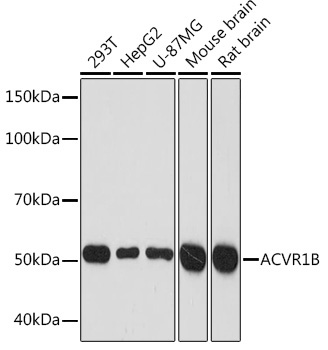
293T, HepG2, U-87MG, Mouse brain, Rat brain
Cellular Localization:
Cell Surface, Cytosol, Plasma Membrane.
Calculated MW:
57kDa
Observed MW:
57kDa
This gene encodes an activin A type IB receptor. Activins are dimeric growth and differentiation factors which belong to the transforming growth factor-beta (TGF-beta) superfamily of structurally related signaling proteins. Activins signal through a heteromeric complex of receptor serine kinases which include at least two type I and two type II receptors. This protein is a type I receptor which is essential for signaling. Mutations in this gene are associated with pituitary tumors. Alternate splicing results in multiple transcript variants. RRID AB_2862987 Gene ID 91 Swiss Prot Synonym ALK4; SKR2; ACTRIB; ACVRLK4; ACVR1B
Purification Method:
Affinity purification
Gene ID:
91
RRID:
AB_2862987
Buffer Information:
Store at -20℃. Avoid freeze / thaw cycles. Buffer: PBS containing 50% glycerol and 0.05% BSA, preserved with proclin300 or sodium azide, pH 7.3.
Western blot analysis of various lysates using ACVR1B Rabbit mAb (CAB2279) at 1:1000 dilution. Secondary antibody: HRP-conjugated Goat anti-Rabbit IgG (H+L) (AS014) at 1:10000 dilution. Lysates/proteins: 25μg per lane. Blocking buffer: 3% nonfat dry milk in TBST. Detection: ECL Basic Kit (AbGn00020). Exposure time: 3min.
Immunohistochemistry analysis of paraffin-embedded Rat testis using ACVR1B Rabbit mAb (CAB2279) at dilution of 1:100 (40x lens). Microwave antigen retrieval performed with 0.01M Tris/EDTA Buffer (pH 9.0) prior to IHC staining.
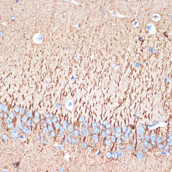
Immunohistochemistry analysis of paraffin-embedded Mouse brain using ACVR1B Rabbit mAb (CAB2279) at dilution of 1:100 (40x lens). Microwave antigen retrieval performed with 0.01M Tris/EDTA Buffer (pH 9.0) prior to IHC staining.
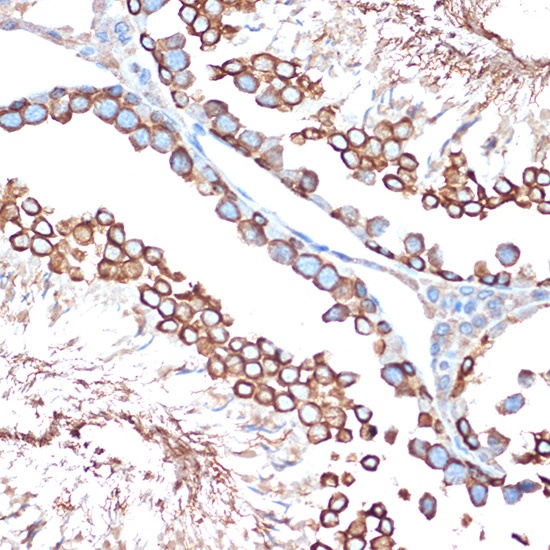
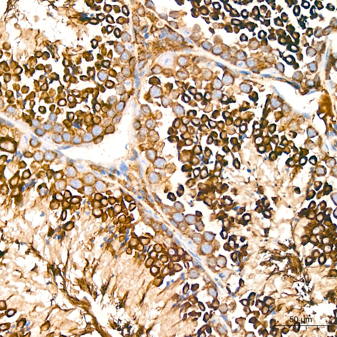
Immunohistochemistry analysis of paraffin-embedded Human cervix cancer tissue using ACVR1B Rabbit mAb (A22347) at a dilution of 1:200 (40x lens). High pressure antigen retrieval performed with 0.01M Citrate buffer (pH 6.0) prior to IHC staining.
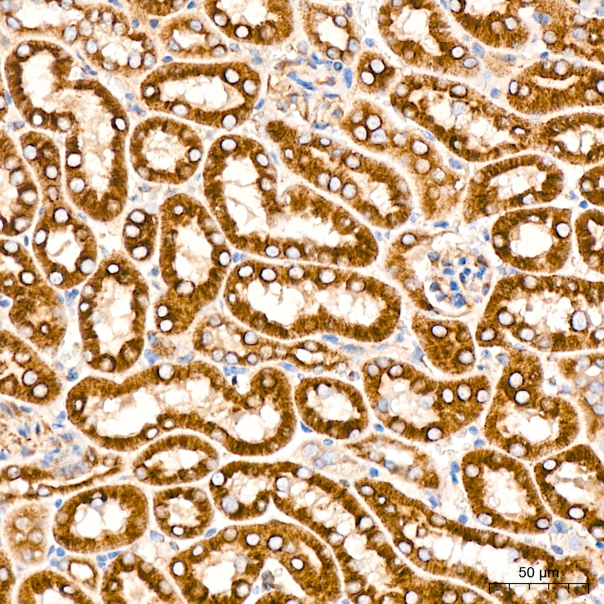
Immunohistochemistry analysis of paraffin-embedded Mouse kidney tissue using ACVR1B Rabbit mAb (A22347) at a dilution of 1:200 (40x lens). High pressure antigen retrieval performed with 0.01M Citrate buffer (pH 6.0) prior to IHC staining.
Immunohistochemistry analysis of paraffin-embedded Mouse testis tissue using ACVR1B Rabbit mAb (A22347) at a dilution of 1:200 (40x lens). High pressure antigen retrieval performed with 0.01M Citrate buffer (pH 6.0) prior to IHC staining.
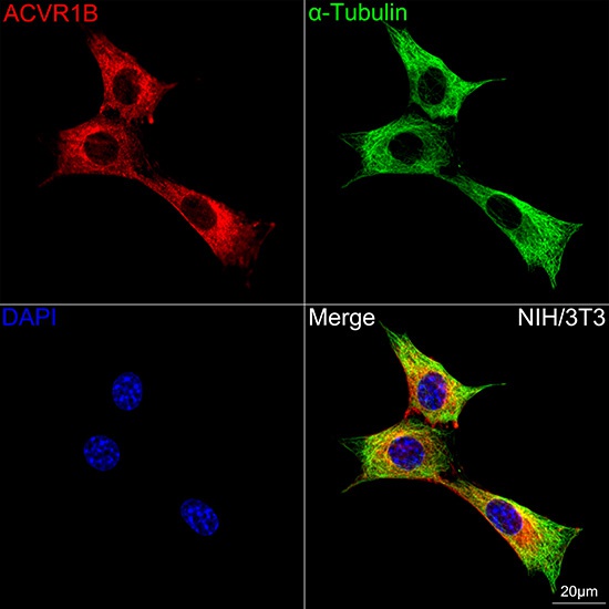
Confocal imaging of NIH/3T3 cells using ACVR1B Rabbit mAb (CAB2279,dilution 1:100) followed by a further incubation with Cy3 Goat Anti-Rabbit IgG (H+L) (AS007,dilution 1:500)(Red).The cells were counterstained with α-Tubulin Mouse mAb (AC012, dilution 1:400) followed by incubation with ABflo® 488-conjugated Goat Anti-Mouse IgG (H+L) Ab (AS076, dilution 1:500) (Green).DAPI was used for nuclear staining (Blue). Objective: 100x.