The AIF1/IBA1 Monoclonal Antibody (CAB19776) is a high-quality antibody developed for reliable detection and analysis of target proteins. Enables actin filament binding activity and calcium ion binding activity. Involved in several processes, including Rac protein signal transduction; actin filament organization; and ruffle assembly. Acts upstream of or within actin filament bundle assembly. Located in several cellular components, including actin filament; phagocytic cup; and ruffle membrane. Is expressed in adrenal cortex; central nervous system; embryo mesenchyme; and retina. Human ortholog(s) of this gene implicated in type 1 diabetes mellitus. Orthologous to human AIF1 (allograft inflammatory factor 1). RRID AB_3711469 Gene ID 11629 Swiss Prot Synonym G1; Iba1; AIF-1; D17H6S50E; AIF1/IBA1
This antibody is validated for use in WB, IHC-P, ELISA, IF-F, IF-P applications and has demonstrated reactivity against Human, Mouse, Rat samples.
Product Name:
AIF1/IBA1 Monoclonal Antibody
SKU:
CAB19776
Size:
100μL, 20μL
Reactivity:
Human, Mouse, Rat
Clone Number:
ARC2301
Conjugate:
Unconjugated
Immunogen:
Synthetic peptide. This information is considered to be commercially sensitive.
Tested Applications:
WBIHC-PELISAIF-FIF-P
Recommended Dilution:
WB
1:1000 - 1:4000
IF-F
1:200 - 1:2000
IF-P
1:200 - 1:2000
IHC-P
1:10000 - 1:40000
ELISA
Recommended starting concentration is 1 μg/mL. Please optimize the concentration based on your specific assay requirements.
Synonyms:
G1, Iba1, AIF-1, D17H6S50E, AIF1/IBA1
Positive Sample:
THP-1, RAW 264.7, Mouse testis, Mouse spleen, Mouse brain, Rat lung, Rat testis, Rat spleen
Cellular Localization:
-
Calculated MW:
17kDa
Observed MW:
17kDa
Enables actin filament binding activity and calcium ion binding activity. Involved in several processes, including Rac protein signal transduction; actin filament organization; and ruffle assembly. Acts upstream of or within actin filament bundle assembly. Located in several cellular components, including actin filament; phagocytic cup; and ruffle membrane. Is expressed in adrenal cortex; central nervous system; embryo mesenchyme; and retina. Human ortholog(s) of this gene implicated in type 1 diabetes mellitus. Orthologous to human AIF1 (allograft inflammatory factor 1). RRID AB_3711469 Gene ID 11629 Swiss Prot Synonym G1; Iba1; AIF-1; D17H6S50E; AIF1/IBA1
Purification Method:
Affinity purification
Gene ID:
11629
RRID:
AB_3711469
Buffer Information:
Store at -20℃. Avoid freeze / thaw cycles. Buffer: PBS with 0.09% sodium azide,0.05% BSA,50% glycerol,pH7.3.
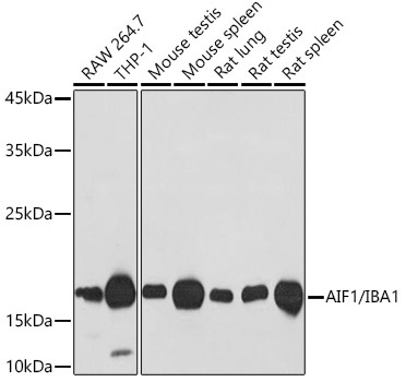
Western blot analysis of various lysates using AIF1/IBA1 Rabbit mAb (CAB19776) at 1:1000 dilution. Secondary antibody: HRP-conjugated Goat anti-Rabbit IgG (H+L) (AS014) at 1:10000 dilution. Lysates/proteins: 25μg per lane. Blocking buffer: 3% nonfat dry milk in TBST. Detection: ECL Basic Kit (AbGn00020). Exposure time: 30s.
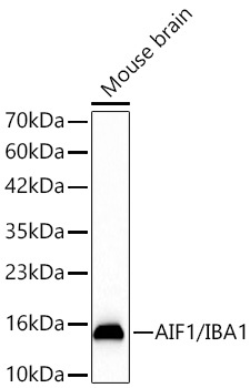
Western blot analysis of lysates from Mouse brain using AIF1/IBA1 Rabbit mAb (CAB19776) at 1:1000 dilution incubated overnight at 4℃. Secondary antibody: HRP-conjugated Goat anti-Rabbit IgG (H+L) (AS014) at 1:10000 dilution. Lysates/proteins: 25 μg per lane. Blocking buffer: 3% nonfat dry milk in TBST. Detection: ECL Basic Kit (AbGn00020). Exposure time: 45s.
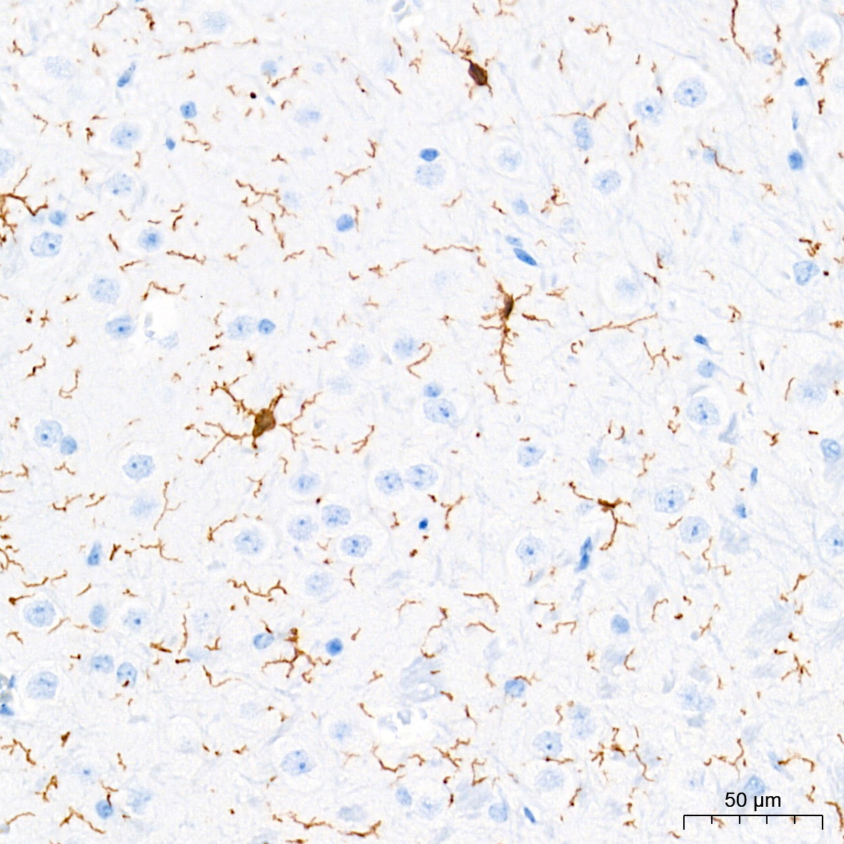
Immunohistochemistry analysis of paraffin-embedded Mouse brain tissue using AIF1/IBA1 Rabbit mAb (CAB19776) at a dilution of 1:20000 (40x lens). High pressure antigen retrieval performed with 0.01M Tris-EDTA Buffer (pH 9.0) prior to IHC staining.
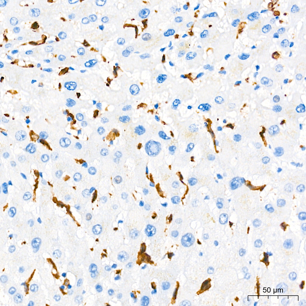
Immunohistochemistry analysis of paraffin-embedded Human liver tissue using AIF1/IBA1 Rabbit mAb (CAB19776) at a dilution of 1:20000 (40x lens). High pressure antigen retrieval performed with 0.01M Tris-EDTA Buffer (pH 9.0) prior to IHC staining.
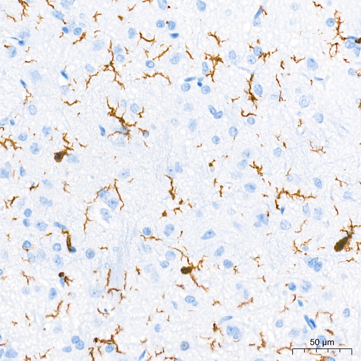
Immunohistochemistry analysis of paraffin-embedded Rat brain tissue using AIF1/IBA1 Rabbit mAb (CAB19776) at a dilution of 1:20000 (40x lens). High pressure antigen retrieval performed with 0.01M Tris-EDTA Buffer (pH 9.0) prior to IHC staining.
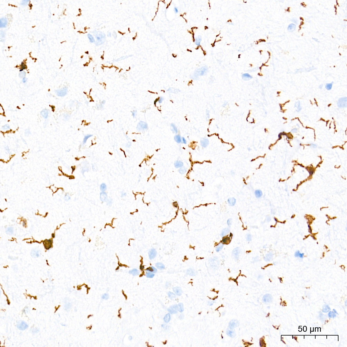
Immunohistochemistry analysis of paraffin-embedded Human brain tissue using AIF1/IBA1 Rabbit mAb (CAB19776) at a dilution of 1:20000 (40x lens). High pressure antigen retrieval performed with 0.01M Tris-EDTA Buffer (pH 9.0) prior to IHC staining.
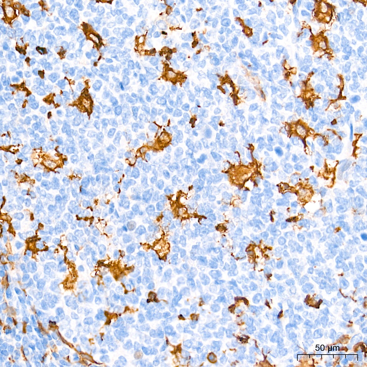
Immunohistochemistry analysis of paraffin-embedded Human tonsil tissue using AIF1/IBA1 Rabbit mAb (CAB19776) at a dilution of 1:20000 (40x lens). High pressure antigen retrieval performed with 0.01M Tris-EDTA Buffer (pH 9.0) prior to IHC staining.
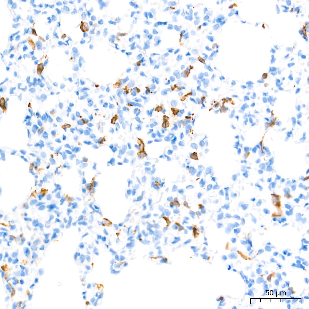
Immunohistochemistry analysis of paraffin-embedded Rat lung tissue using AIF1/IBA1 Rabbit mAb (CAB19776) at a dilution of 1:20000 (40x lens). High pressure antigen retrieval performed with 0.01M Tris-EDTA Buffer (pH 9.0) prior to IHC staining.
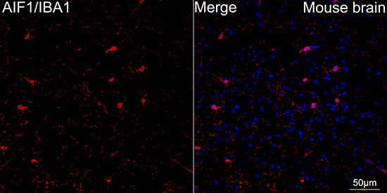
Confocal imaging of paraffin-embedded Mouse brain using AIF1/IBA1 Rabbit mAb (CAB19776, dilution 1:200) followed by a further incubation with Cy3 Goat Anti-Rabbit IgG (H+L) (AS007, dilution 1:500) (Red). DAPI was used for nuclear staining (Blue). Objective: 40x.Perform microwave antigen retrieval with 0.01M citrate buffer (pH 6.0) prior to IF staining.
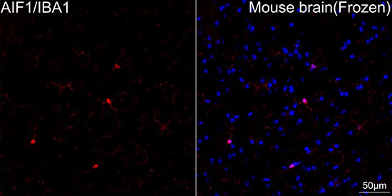
Confocal imaging of frozen sections Mouse brain tissue using AIF1/IBA1 Rabbit mAb (CAB19776, dilution 1:200) followed by a further incubation with Cy3 Goat Anti-Rabbit IgG (H+L) (AS007, dilution 1:500) (Red). DAPI was used for nuclear staining (Blue). Microwave antigen retrieval performed with 0.01M Citrate Buffer (pH 6.0) prior to IF staining. Objective: 40x.
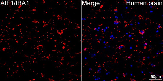
Confocal imaging of paraffin-embedded Human brain tissue using AIF1/IBA1 Rabbit mAb (CAB19776, dilution 1:200) followed by a further incubation with Cy3 Goat Anti-Rabbit IgG (H+L) (AS007, dilution 1:500) (Red). DAPI was used for nuclear staining (Blue). High pressure antigen retrieval performed with 0.01M Citrate Buffer (pH 6.0) prior to IF staining. Objective: 40x.