The alpha-Tubulin Monoclonal Antibody (CAB6830) is a high-quality antibody developed for reliable detection and analysis of target proteins. This polyclonal antibody, raised in rabbits, specifically targets alpha tubulin, a vital component of microtubules involved in cell structure and transport processes.This antibody is highly reactive with human samples and is validated for use in Western blot applications, allowing for accurate detection and analysis of alpha tubulin expression. Its specificity towards alpha tubulin makes it an essential tool for researchers investigating cell division, intracellular transport, and cell motility.
This antibody is validated for use in WB, IHC-P, IF/ICC, IP, ELISA applications and has demonstrated reactivity against Human, Mouse, Rat samples.
Product Name:
alpha-Tubulin Monoclonal Antibody
SKU:
CAB6830
Size:
20μL, 100μL
Reactivity:
Human, Mouse, Rat
Clone Number:
ARC2486
Conjugate:
Unconjugated
Immunogen:
Synthetic peptide. This information is considered to be commercially sensitive.
Microtubules of the eukaryotic cytoskeleton perform essential and diverse functions and are composed of a heterodimer of alpha and beta tubulin. The genes encoding these microtubule constituents are part of the tubulin superfamily, which is composed of six distinct families. Genes from the alpha, beta and gamma tubulin families are found in all eukaryotes. The alpha and beta tubulins represent the major components of microtubules, while gamma tubulin plays a critical role in the nucleation of microtubule assembly. There are multiple alpha and beta tubulin genes and they are highly conserved among and between species. This gene encodes an alpha tubulin that is a highly conserved homolog of a rat testis-specific alpha tubulin. Alternatively spliced transcript variants encoding different isoforms have been found for this gene.
Purification Method
Affinity purification
Gene ID
7277
RRID
AB_2863541
Buffer Information
Store at -20℃. Avoid freeze / thaw cycles. Buffer: PBS containing 50% glycerol and 0.05% BSA, preserved with proclin300 or sodium azide, pH 7.3.
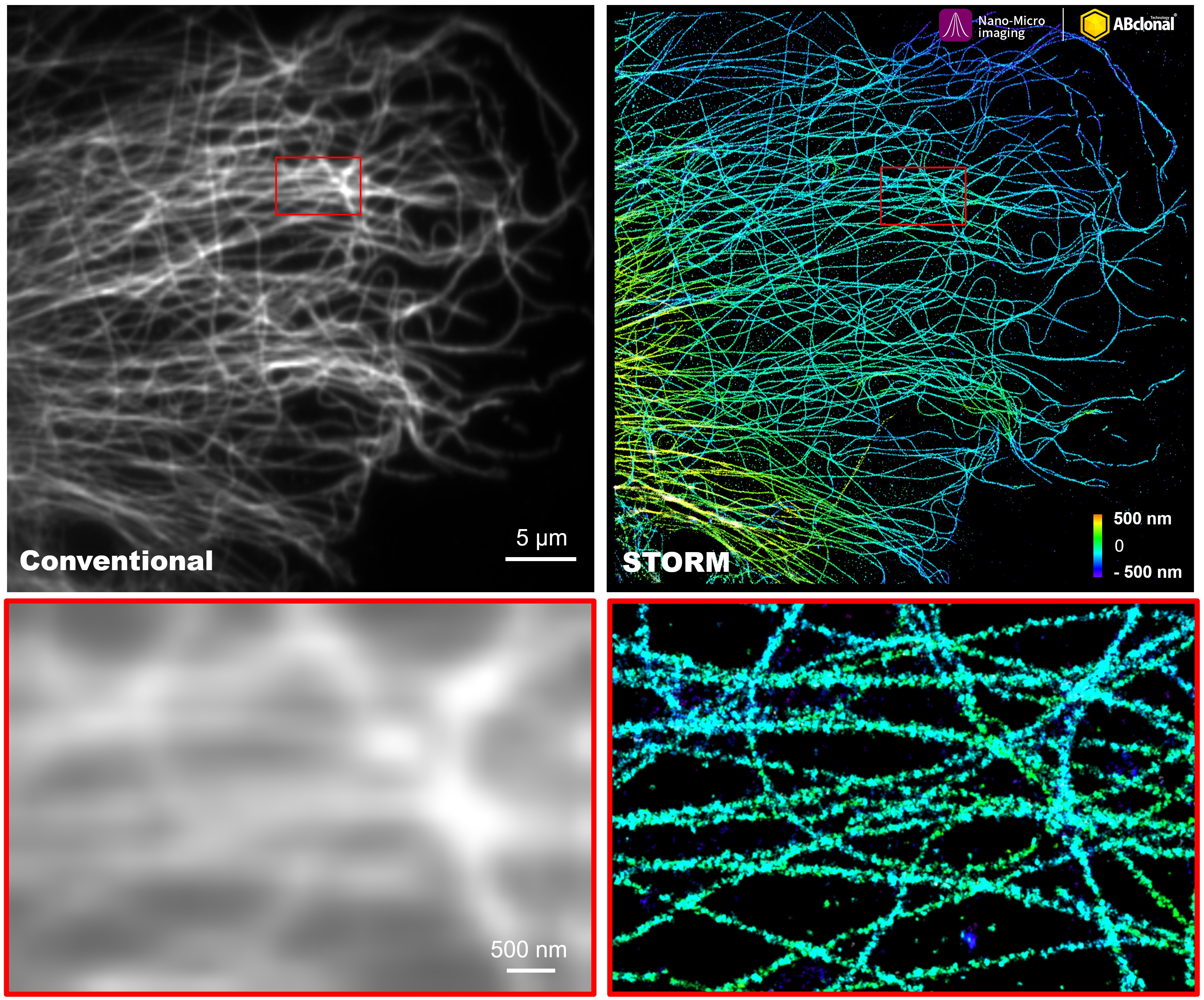
The STORM super-resolution (SR) imaging of COS7 cells using α-Tubulin Rabbit mAb (CAB6830, ABclonal) at dilution of 1:50 with 3% paraformaldehyde (PFA) +0.1% glutaraldehyde (GA) fixation. The immunostaining was performed by Full Automatic Immunofluorescence Workflow System (Workflow Ultra300, Nano-Micro imaging, China). Image was performed with Single-Molecule Localization Super-Resolution Microscopy (STORM Ultra300, Nano-Micro imaging, China). We acknowledge Nano-Micro imaging Biotechnology Co., Ltd. in SR image processing and kindly providing this image.
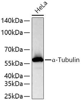
Western blot analysis of lysates from HeLa cells using α-Tubulin Rabbit mAb (CAB6830) at 1:10000 dilution incubated overnight at 4℃. Secondary antibody: HRP-conjugated Goat anti-Rabbit IgG (H+L) (CABS014) at 1:10000 dilution. Lysates/proteins: 25 μg per lane. Blocking buffer: 3% nonfat dry milk in TBST. Detection: ECL Basic Kit (AbGn00020). Exposure time: 10 s.
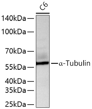
Western blot analysis of lysates from C6 cells using α-Tubulin Rabbit mAb (CAB6830) at 1:10000 dilution incubated overnight at 4℃. Secondary antibody: HRP-conjugated Goat anti-Rabbit IgG (H+L) (CABS014) at 1:10000 dilution. Lysates/proteins: 25 μg per lane. Blocking buffer: 3% nonfat dry milk in TBST. Detection: ECL Basic Kit (AbGn00020). Exposure time: 45 s.
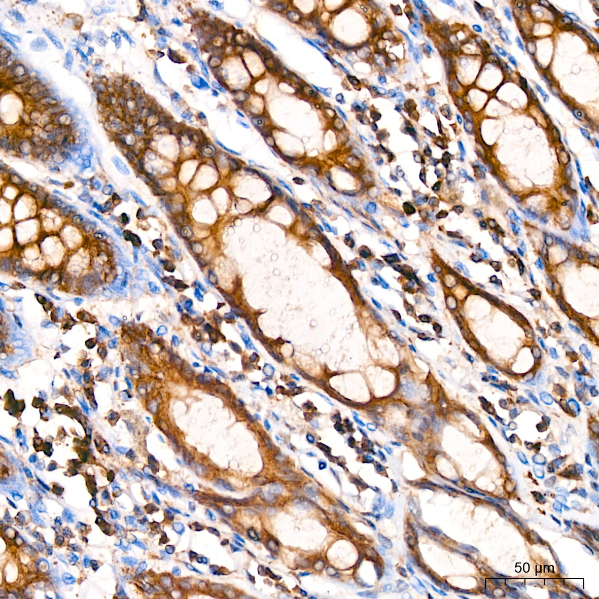
Immunohistochemistry analysis of paraffin-embedded Human colon using α-Tubulin Rabbit mAb (CAB6830) at dilution of 1:200 (40x lens). High pressure antigen retrieval performed with 0.01M Citrate buffer (pH 6.0) prior to IHC staining.
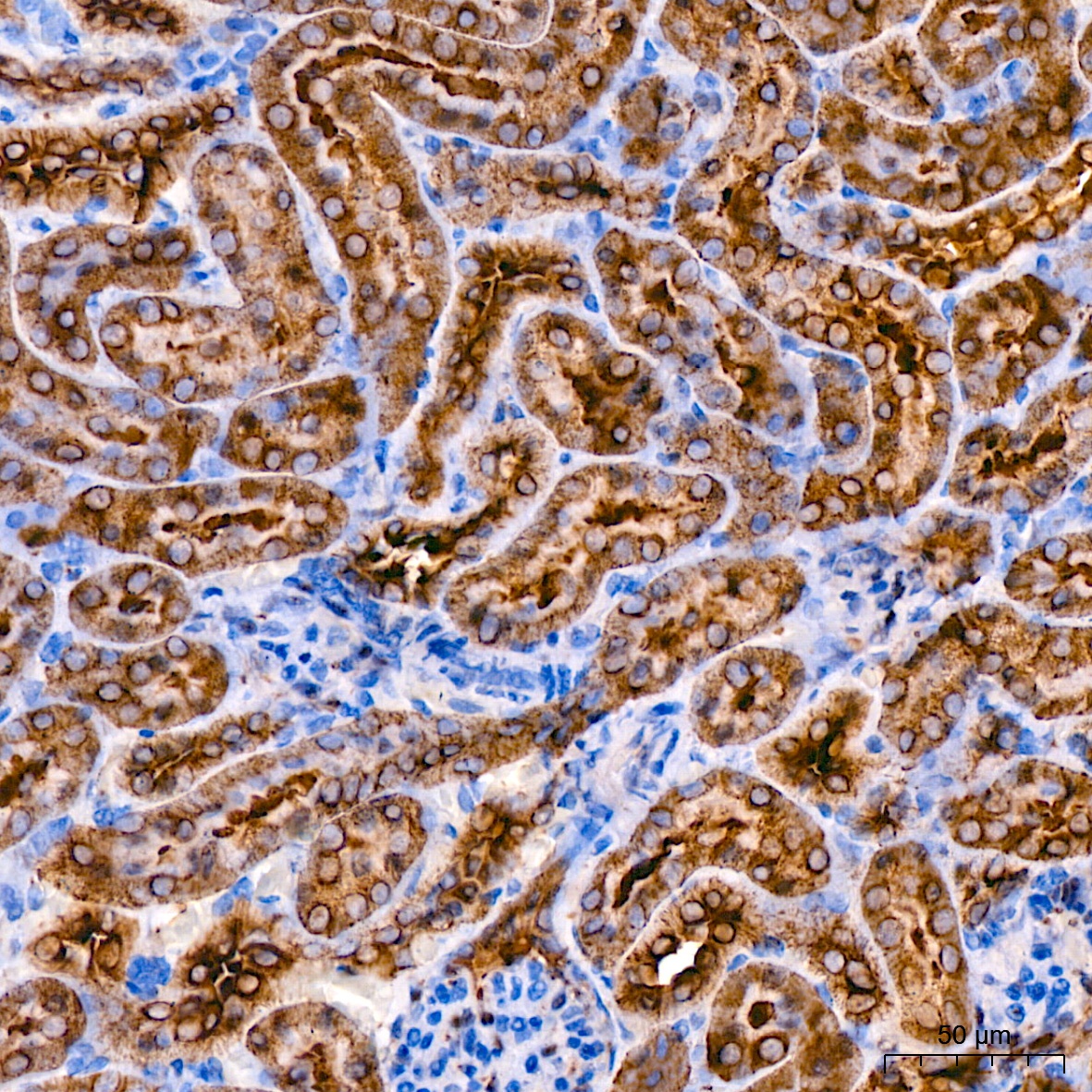
Immunohistochemistry analysis of paraffin-embedded Mouse kidney using α-Tubulin Rabbit mAb (CAB6830) at dilution of 1:200 (40x lens). High pressure antigen retrieval performed with 0.01M Citrate buffer (pH 6.0) prior to IHC staining.
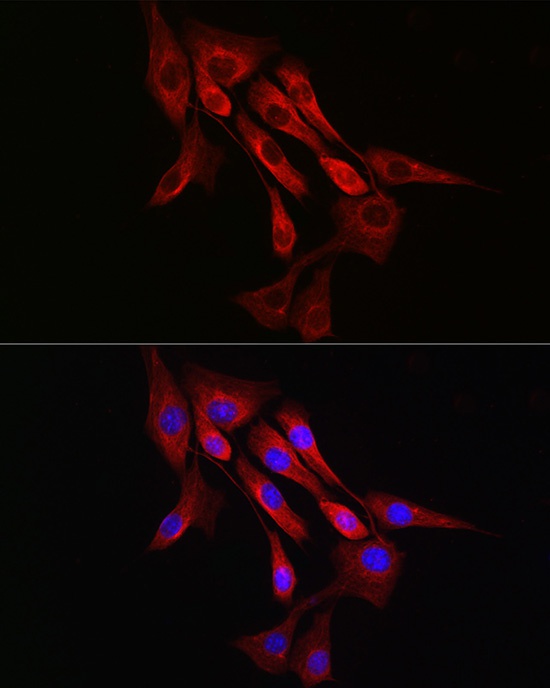
Immunofluorescence analysis of NIH/3T3 cells using α-Tubulin Rabbit mAb (CAB6830) at dilution of 1:100 (40x lens). Secondary antibody: Cy3-conjugated Goat anti-Rabbit IgG (H+L) (CABS007) at 1:500 dilution. Blue: DAPI for nuclear staining.
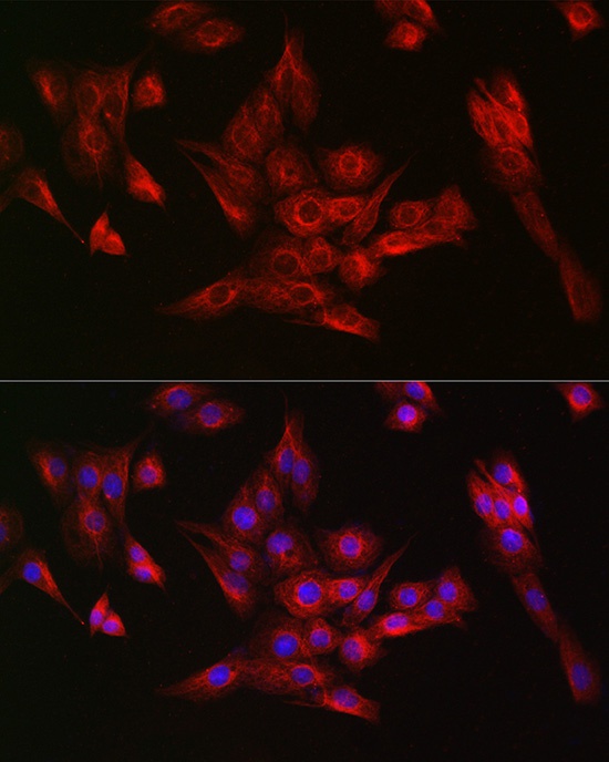
Immunofluorescence analysis of PC-12 cells using α-Tubulin Rabbit mAb (CAB6830) at dilution of 1:100 (40x lens). Secondary antibody: Cy3-conjugated Goat anti-Rabbit IgG (H+L) (CABS007) at 1:500 dilution. Blue: DAPI for nuclear staining.
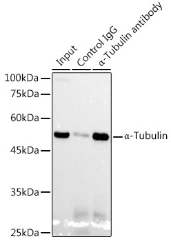
Immunoprecipitation analysis of 300 μg extracts from HeLa cells using 3 μg α-Tubulin Rabbit mAb (CAB6830). Western blot was performed from the immunoprecipitate using α-Tubulin Rabbit mAb (CAB6830) at a dilution of 1:1000.


")


