The ANP32B Monoclonal Antibody (CAB3489) is a high-quality antibody developed for reliable detection and analysis of target proteins. This antibody, produced in rabbits, exhibits high reactivity with human samples and has been validated for use in techniques such as Western blotting. By binding to the ANP32B protein, this antibody enables precise detection and analysis in different cell types, making it an ideal choice for studies in cell biology and cancer research.ANP32B, also known as Acidic leucine-rich nuclear phosphoprotein 32 family member B, plays a crucial role in regulating gene expression, cell cycle progression, and apoptosis.
This antibody is validated for use in WB, IHC-P, IF/ICC, ELISA, IF-P applications and has demonstrated reactivity against Human, Mouse, Rat samples.
Product Name:
ANP32B Monoclonal Antibody
SKU:
CAB3489
Size:
20μL, 100μL
Reactivity:
Human, Mouse, Rat
Clone Number:
ARC2014
Conjugate:
Unconjugated
Immunogen:
Synthetic peptide. This information is considered to be commercially sensitive.
Recommended starting concentration is 1 μg/mL. Please optimize the concentration based on your specific assay requirements.
Synonyms:
APRIL, SSP29, PHAPI2, ANP32B
Positive Sample:
A549, PC-3, RAW264.7, U-937, Mouse testis
Cellular Localization:
Cytoplasm, Nucleus.
Calculated MW:
29kDa
Observed MW:
28-31kDa
Enables RNA polymerase binding activity and histone binding activity. Involved in several processes, including activation of cysteine-type endopeptidase activity involved in apoptotic process; nucleosome assembly; and positive regulation of protein export from nucleus. Located in cytoplasm and nucleoplasm. Colocalizes with nucleolus.
Purification Method
Affinity purification
Gene ID
10541
RRID
AB_2863071
Buffer Information
Store at -20℃. Avoid freeze / thaw cycles. Buffer: PBS containing 50% glycerol and 0.05% BSA, preserved with proclin300 or sodium azide, pH 7.3.
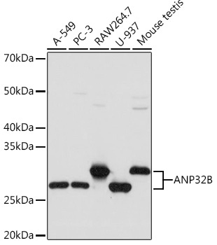
Western blot analysis of various lysates using ANP32B Rabbit mAb (CAB3489) at 1:1000 dilution. Secondary antibody: HRP-conjugated Goat anti-Rabbit IgG (H+L) (CABS014) at 1:10000 dilution. Lysates/proteins: 25μg per lane. Blocking buffer: 3% nonfat dry milk in TBST. Detection: ECL Basic Kit (AbGn00020). Exposure time: 3s.
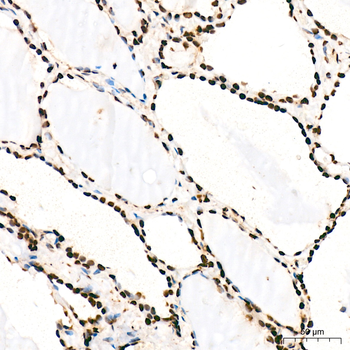
Immunohistochemistry analysis of paraffin-embedded Human thyroid tissue using ANP32B Rabbit mAb (CAB3489) at a dilution of 1:200 (40x lens). High pressure antigen retrieval was performed with 0.01 M citrate buffer (pH 6.0) prior to IHC staining.
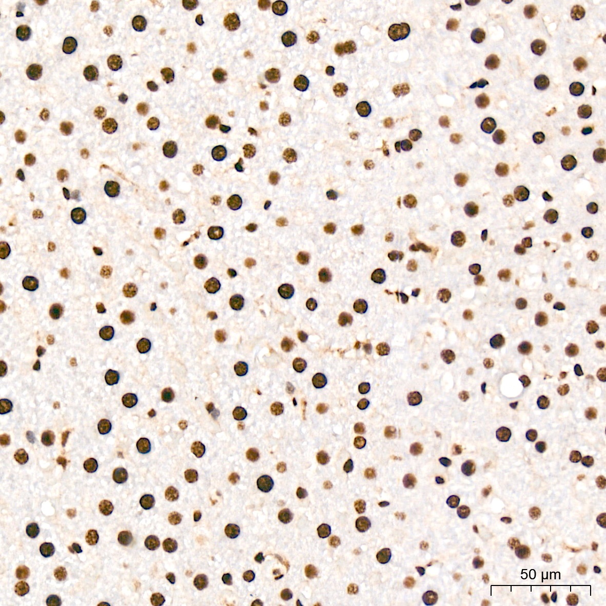
Immunohistochemistry analysis of paraffin-embedded Mouse liver tissue using ANP32B Rabbit mAb (CAB3489) at a dilution of 1:200 (40x lens). High pressure antigen retrieval was performed with 0.01 M citrate buffer (pH 6.0) prior to IHC staining.
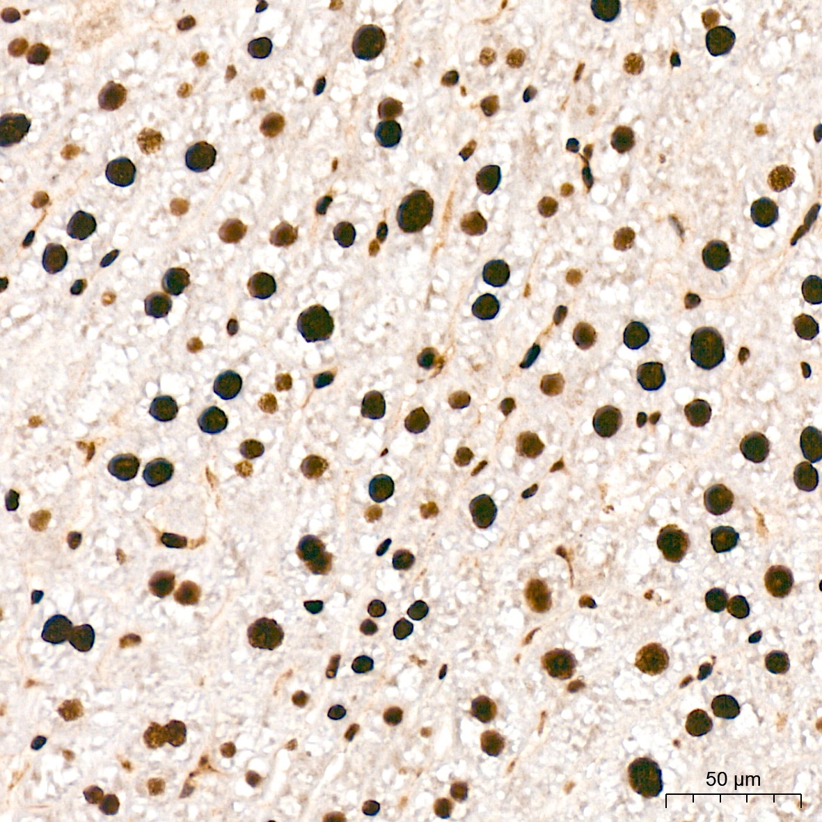
Immunohistochemistry analysis of paraffin-embedded Rat liver tissue using ANP32B Rabbit mAb (CAB3489) at a dilution of 1:200 (40x lens). High pressure antigen retrieval was performed with 0.01 M citrate buffer (pH 6.0) prior to IHC staining.
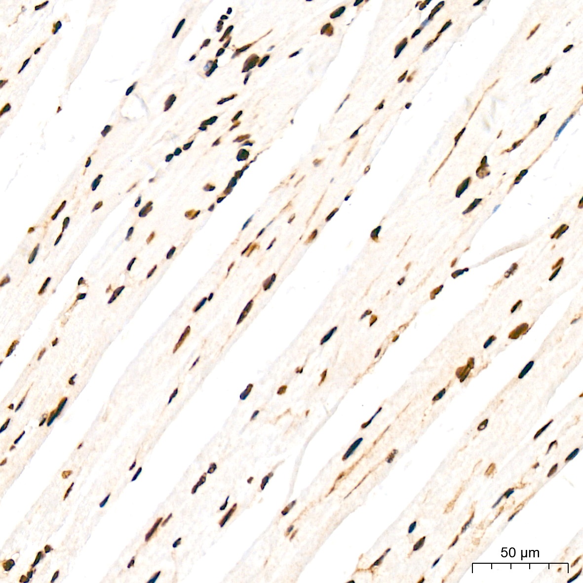
Immunohistochemistry analysis of paraffin-embedded Mouse heart tissue using ANP32B Rabbit mAb (CAB3489) at a dilution of 1:200 (40x lens). High pressure antigen retrieval was performed with 0.01 M citrate buffer (pH 6.0) prior to IHC staining.
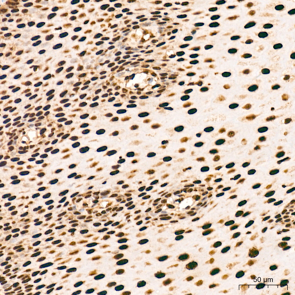
Immunohistochemistry analysis of paraffin-embedded Human esophagus tissue using ANP32B Rabbit mAb (CAB3489) at a dilution of 1:200 (40x lens). High pressure antigen retrieval was performed with 0.01 M citrate buffer (pH 6.0) prior to IHC staining.
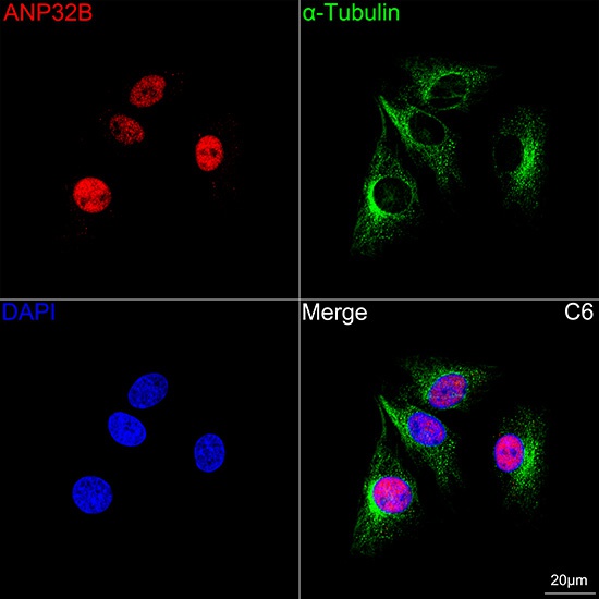
Confocal imaging of C6 cells using ANP32B Rabbit mAb (CAB3489, dilution 1:200) followed by a further incubation with Cy3 Goat Anti-Rabbit IgG (H+L) (CABS007, dilution 1:500) (Red). The cells were counterstained with α-Tubulin Mouse mAb (AC012, dilution 1:400) followed by incubation with ABflo® 488-conjugated Goat Anti-Mouse IgG (H+L) Ab (CABS076, dilution 1:500) (Green). DAPI was used for nuclear staining (Blue). Objective: 100x.
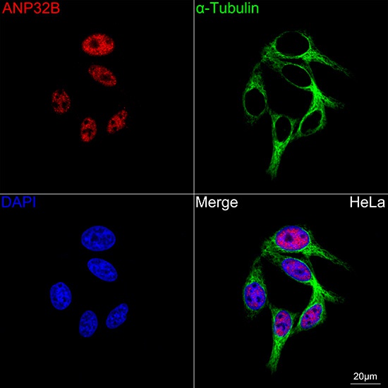
Confocal imaging of HeLa cells using ANP32B Rabbit mAb (CAB3489, dilution 1:200) followed by a further incubation with Cy3 Goat Anti-Rabbit IgG (H+L) (CABS007, dilution 1:500) (Red). The cells were counterstained with α-Tubulin Mouse mAb (AC012, dilution 1:400) followed by incubation with ABflo® 488-conjugated Goat Anti-Mouse IgG (H+L) Ab (CABS076, dilution 1:500) (Green). DAPI was used for nuclear staining (Blue). Objective: 100x.
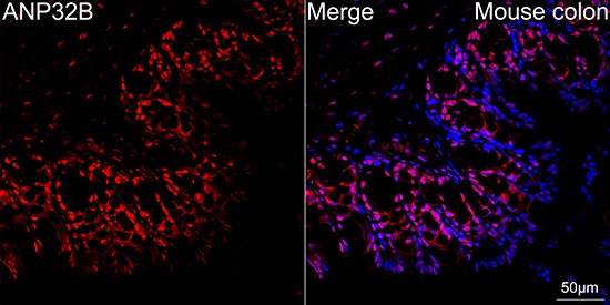
Confocal imaging of paraffin-embedded Mouse colon tissue using ANP32B Rabbit mAb (CAB3489, dilution 1:200) followed by a further incubation with Cy3 Goat Anti-Rabbit IgG (H+L) (CABS007, dilution 1:500) (Red). DAPI was used for nuclear staining (Blue). Objective: 40x. Perform high pressure antigen retrieval with 0.01 M citrate buffer (pH 6.0) prior to IF staining.