
Description
ATP6V1G2 Antibody (CAB20163)
The ATP6V1G2 Antibody (CAB20163) is a high-quality antibody developed for reliable detection and analysis of target proteins. This antibody, raised in rabbits, exhibits high reactivity with human samples and has been validated for use in Western blot applications.ATP6V1G2 is crucial for maintaining the acidic pH of various organelles and regulating cell growth and proliferation. Dysregulation of ATP6V1G2 has been linked to diseases such as cancer and osteopetrosis, making it a target of interest in biomedical research.
This antibody is validated for use in WB, IHC-P, ELISA applications and has demonstrated reactivity against Human, Mouse, Rat samples.
| Product Name: | ATP6V1G2 Antibody |
| SKU: | CAB20163 |
| Size: | 20μL, 100μL |
| Reactivity: | Human, Mouse, Rat |
| Conjugate: | Unconjugated |
| Immunogen: | Recombinant protein (or fragment).This information is considered to be commercially sensitive. | ||||||
| Sequence: | AQME VEQY RRER EHEF QSKQ QAAM GSQG NLSA EVEQ ATRR QVQG MQSS QQRN RERV LAQL L | ||||||
| Tested Applications: | WB IHC-P ELISA | ||||||
| Recommended Dilution: |
| ||||||
| Synonyms: | NG38, ATP6G, VMA10, ATP6G2, ATP6V1G2 |
| Positive Sample: | SH-SY5Y, Mouse brain, Rat brain, Mouse eye |
| Cellular Localization: | Cytosol, Extrinsic Component Of Synaptic Vesicle Membrane. |
| Calculated MW: | 14kDa |
| Observed MW: | 14kDa |
This gene encodes a component of vacuolar ATPase (V-ATPase), a multisubunit enzyme that mediates acidification of intracellular compartments of eukaryotic cells. V-ATPase dependent acidification is necessary for such intracellular processes as protein sorting, zymogen activation, receptor-mediated endocytosis, and synaptic vesicle proton gradient generation. V-ATPase is composed of a cytosolic V1 domain and a transmembrane V0 domain. The V1 domain consists of three A and three B subunits, two G subunits plus the C, D, E, F, and H subunits. The V1 domain contains the ATP catalytic site. The V0 domain consists of five different subunits: a, c, c', c'', and d. Additional isoforms of many of the V1 and V0 subunit proteins are encoded by multiple genes or alternatively spliced transcript variants. This encoded protein is one of three V1 domain G subunit proteins. This gene had previous gene symbols of ATP6G and ATP6G2. Alternatively spliced transcript variants encoding different isoforms have been described. Read-through transcription also exists between this gene and the downstream DEAD (Asp-Glu-Ala-Asp) box polypeptide 39B (DDX39B) gene.
| Purification Method | Affinity purification |
| Gene ID | 534 |
| RRID | AB_2862950 |
| Buffer Information | Store at -20℃. Avoid freeze / thaw cycles. Buffer: PBS containing 50% glycerol, preserved with proclin300 or sodium azide, pH 7.3. |
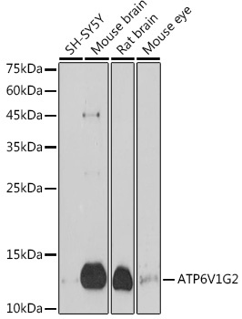 | Western blot analysis of various lysates using ATP6V1G2 Rabbit pAb (CAB20163) at 1:1000 dilution. Secondary antibody: HRP-conjugated Goat anti-Rabbit IgG (H+L) (CABS014) at 1:10000 dilution. Lysates/proteins: 25μg per lane. Blocking buffer: 3% nonfat dry milk in TBST. Detection: ECL Basic Kit (AbGn00020). Exposure time: 180s. |
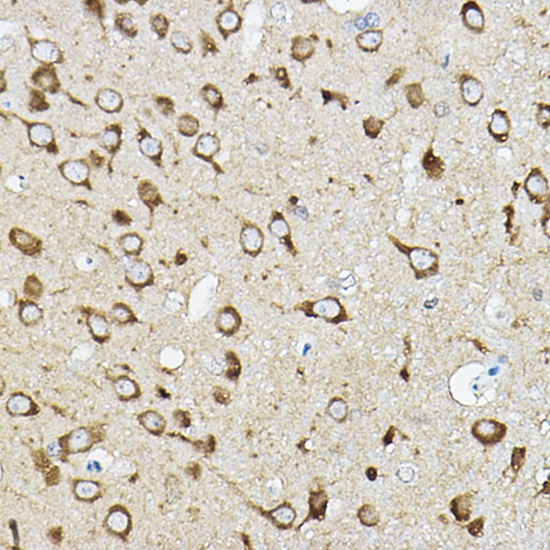 | Immunohistochemistry analysis of paraffin-embedded Rat brain using ATP6V1G2 Rabbit pAb (CAB20163) at dilution of 1:50 (40x lens). High pressure antigen retrieval performed with 0.01M Citrate buffer (pH 6.0) prior to IHC staining. |


