The Cadherin 16 Monoclonal Antibody (CAB19644) is a high-quality antibody developed for reliable detection and analysis of target proteins. This antibody, produced in rabbits, is highly specific for human samples and has been validated for use in Western blot applications. By binding to Cadherin 16, this antibody allows for the detection and analysis of this protein in a variety of cell types, making it ideal for investigations in developmental biology and cancer research.
This antibody is validated for use in WB, ELISA, IF-P applications and has demonstrated reactivity against Human, Mouse samples.
Product Name:
Cadherin 16 Monoclonal Antibody
SKU:
CAB19644
Size:
20μL, 100μL
Reactivity:
Human, Mouse
Clone Number:
ARC2212
Conjugate:
Unconjugated
Immunogen:
Recombinant protein (or fragment).This information is considered to be commercially sensitive.
This gene is a member of the cadherin superfamily, genes encoding calcium-dependent, membrane-associated glycoproteins. Mapped to a previously identified cluster of cadherin genes on chromosome 16q22.1, the gene localizes with superfamily members CDH1, CDH3, CDH5, CDH8 and CDH11. The protein consists of an extracellular domain containing 6 cadherin domains, a transmembrane region and a truncated cytoplasmic domain but lacks the prosequence and tripeptide HAV adhesion recognition sequence typical of most classical cadherins. Expression is exclusively in kidney, where the protein functions as the principal mediator of homotypic cellular recognition, playing a role in the morphogenic direction of tissue development. Alternatively spliced transcript variants encoding distinct isoforms have been identified.
Purification Method
Affinity purification
Gene ID
1014
Buffer Information
Store at -20℃. Avoid freeze / thaw cycles. Buffer: PBS containing 50% glycerol and 0.05% BSA, preserved with proclin300 or sodium azide, pH 7.3.
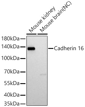
Western blot analysis of various lysates using Cadherin 16 Rabbit mAb (CAB19644) at 1:1000 dilution incubated at room temperature for 1.5 hours. Secondary antibody: HRP-conjugated Goat anti-Rabbit IgG (H+L) (CABS014) at 1:10000 dilution. Lysates/proteins: 25 μg per lane. Blocking buffer: 3% nonfat dry milk in TBST. Detection: ECL Basic Kit (AbGn00020). Negative control (NC): Mouse brain. Exposure time: 5 s.
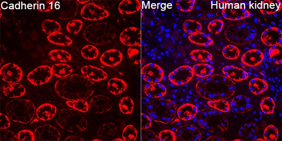
Confocal imaging of paraffin-embedded Human kidney tissue using Cadherin 16 Rabbit mAb (CAB19644, dilution 1:200) followed by a further incubation with Cy3 Goat Anti-Rabbit IgG (H+L) (CABS007, dilution 1:500) (Red). DAPI was used for nuclear staining (Blue). High pressure antigen retrieval performed with 0.01M Citrate Buffer (pH 6.0) prior to IF staining. Objective: 40x.