The CD79B Monoclonal Antibody (CAB4362) is a high-quality antibody developed for reliable detection and analysis of target proteins. The B lymphocyte antigen receptor is a multimeric complex that includes the antigen-specific component, surface immunoglobulin (Ig). Surface Ig non-covalently associates with two other proteins, Ig-alpha and Ig-beta, which are necessary for expression and function of the B-cell antigen receptor. This gene encodes the Ig-beta protein of the B-cell antigen component. Alternatively spliced transcript variants encoding different isoforms have been described.
This antibody is validated for use in WB, IHC-P, ELISA, IF-P applications and has demonstrated reactivity against Human, Mouse, Rat samples.
Product Name:
CD79B Monoclonal Antibody
SKU:
CAB4362
Size:
100μL, 20μL
Reactivity:
Human, Mouse, Rat
Clone Number:
ARC52688
Conjugate:
Unconjugated
Immunogen:
Synthetic peptide. This information is considered to be commercially sensitive.
Tested Applications:
WBIHC-PELISAIF-P
Recommended Dilution:
WB
1:2000 - 1:4000
IF-P
1:500 - 1:1000
IHC-P
1:500 - 1:2000
ELISA
Recommended starting concentration is 1 μg/mL. Please optimize the concentration based on your specific assay requirements.
Synonyms:
B29, IGB, AGM6, Igbeta, CD79B
Positive Sample:
Daudi, Raji, Rat spleen
Cellular Localization:
External Side Of Plasma Membrane, Extracellular Exosome, Plasma Membrane.
Calculated MW:
26kDa
Observed MW:
30-50kDa
The B lymphocyte antigen receptor is a multimeric complex that includes the antigen-specific component, surface immunoglobulin (Ig). Surface Ig non-covalently associates with two other proteins, Ig-alpha and Ig-beta, which are necessary for expression and function of the B-cell antigen receptor. This gene encodes the Ig-beta protein of the B-cell antigen component. Alternatively spliced transcript variants encoding different isoforms have been described.
Purification Method
Affinity purification
Gene ID
974
Buffer Information
Store at -20℃. Avoid freeze / thaw cycles. Buffer: PBS containing 50% glycerol and 0.05% BSA, preserved with proclin300 or sodium azide, pH 7.3.
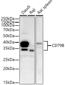
Western blot analysis of various lysates, using CD79B Rabbit mAb (CAB4362) at 1:3600 dilution. Secondary antibody: HRP-conjugated Goat anti-Rabbit IgG (H+L) (AS014) at 1:10000 dilution. Lysates/proteins: 25μg per lane. Blocking buffer: 3% nonfat dry milk in TBST. Detection: ECL Basic Kit (AbGn00020). Exposure time: 60s.
Immunohistochemistry analysis of paraffin-embedded Human tonsil tissue using CD79B Rabbit mAb (CAB4362) at a dilution of 1:2000 (40x lens). High pressure antigen retrieval performed with 0.01M Tris-EDTA Buffer (pH 9.0) prior to IHC staining.
Confocal imaging of paraffin-embedded Human spleen using CD79B Rabbit mAb (CAB4362,dilution 1:1000) followed by a further incubation with Cy3 Goat Anti-Rabbit IgG (H+L) (AS007,dilution 1:500)(Red).DAPI was used for nuclear staining (Blue). Objective: 40x.Perform high pressure antigen retrieval with 0.01M citrate buffer (pH 6.0) prior to IF staining.
Confocal imaging of paraffin-embedded mouse spleen using CD79B Rabbit mAb (CAB4362,dilution 1:1000) followed by a further incubation with Cy3 Goat Anti-Rabbit IgG (H+L) (AS007,dilution 1:500)(Red).DAPI was used for nuclear staining (Blue). Objective: 40x.Perform high pressure antigen retrieval with 0.01M citrate buffer (pH 6.0) prior to IF staining.