The DCR2 Monoclonal Antibody (CAB3581) is a high-quality antibody developed for reliable detection and analysis of target proteins. The protein encoded by this gene is a member of the TNF-receptor superfamily. This receptor contains an extracellular TRAIL-binding domain, a transmembrane domain, and a truncated cytoplamic death domain. This receptor does not induce apoptosis, and has been shown to play an inhibitory role in TRAIL-induced cell apoptosis. RRID Gene ID 8793 Swiss Prot Synonym DCR2; CD264; TRUNDD; TRAILR4; TRAIL-R4
This antibody is validated for use in WB, IHC-P, ELISA applications and has demonstrated reactivity against Human, Mouse, Rat samples.
Product Name:
DCR2 Monoclonal Antibody
SKU:
CAB3581
Size:
100μL, 20μL
Reactivity:
Human, Mouse, Rat
Clone Number:
ARC2052
Conjugate:
Unconjugated
Immunogen:
Synthetic peptide. This information is considered to be commercially sensitive.
Tested Applications:
WBIHC-PELISA
Recommended Dilution:
WB
1:1000 - 1:6000
IHC-P
1:50 - 1:200
ELISA
Recommended starting concentration is 1 μg/mL. Please optimize the concentration based on your specific assay requirements.
Synonyms:
DCR2, CD264, TRUNDD, TRAILR4, TRAIL-R4
Positive Sample:
Mouse liver, Mouse kidney, Rat liver, Rat kidney
Cellular Localization:
Cell Surface, Plasma Membrane.
Calculated MW:
42kDa
Observed MW:
50kDa
The protein encoded by this gene is a member of the TNF-receptor superfamily. This receptor contains an extracellular TRAIL-binding domain, a transmembrane domain, and a truncated cytoplamic death domain. This receptor does not induce apoptosis, and has been shown to play an inhibitory role in TRAIL-induced cell apoptosis. RRID Gene ID 8793 Swiss Prot Synonym DCR2; CD264; TRUNDD; TRAILR4; TRAIL-R4
Purification Method:
Affinity purification
Gene ID:
8793
RRID:
-
Buffer Information:
Store at -20℃. Avoid freeze / thaw cycles. Buffer: PBS containing 50% glycerol and 0.05% BSA, preserved with proclin300 or sodium azide, pH 7.3.
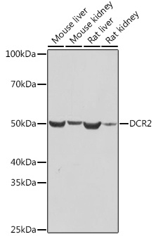
Western blot analysis of various lysates using DCR2 Rabbit mAb (CAB3581) at 1:1000 dilution. Secondary antibody: HRP-conjugated Goat anti-Rabbit IgG (H+L) (AS014) at 1:10000 dilution. Lysates/proteins: 25μg per lane. Blocking buffer: 3% nonfat dry milk in TBST. Detection: ECL Basic Kit (AbGn00020). Exposure time: 10s.
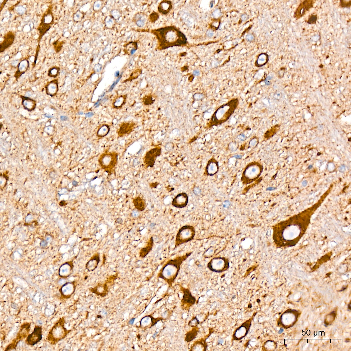
Immunohistochemistry analysis of paraffin-embedded Rat brain tissue using DCR2 Rabbit mAb (CAB3581) at a dilution of 1:200 (40x lens). High pressure antigen retrieval was performed with 0.01 M citrate buffer (pH 6.0) prior to IHC staining.
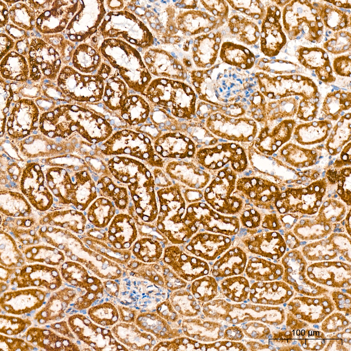
Immunohistochemistry analysis of paraffin-embedded Mouse kidney tissue using DCR2 Rabbit mAb (CAB3581) at a dilution of 1:200 (40x lens). High pressure antigen retrieval was performed with 0.01 M citrate buffer (pH 6.0) prior to IHC staining.
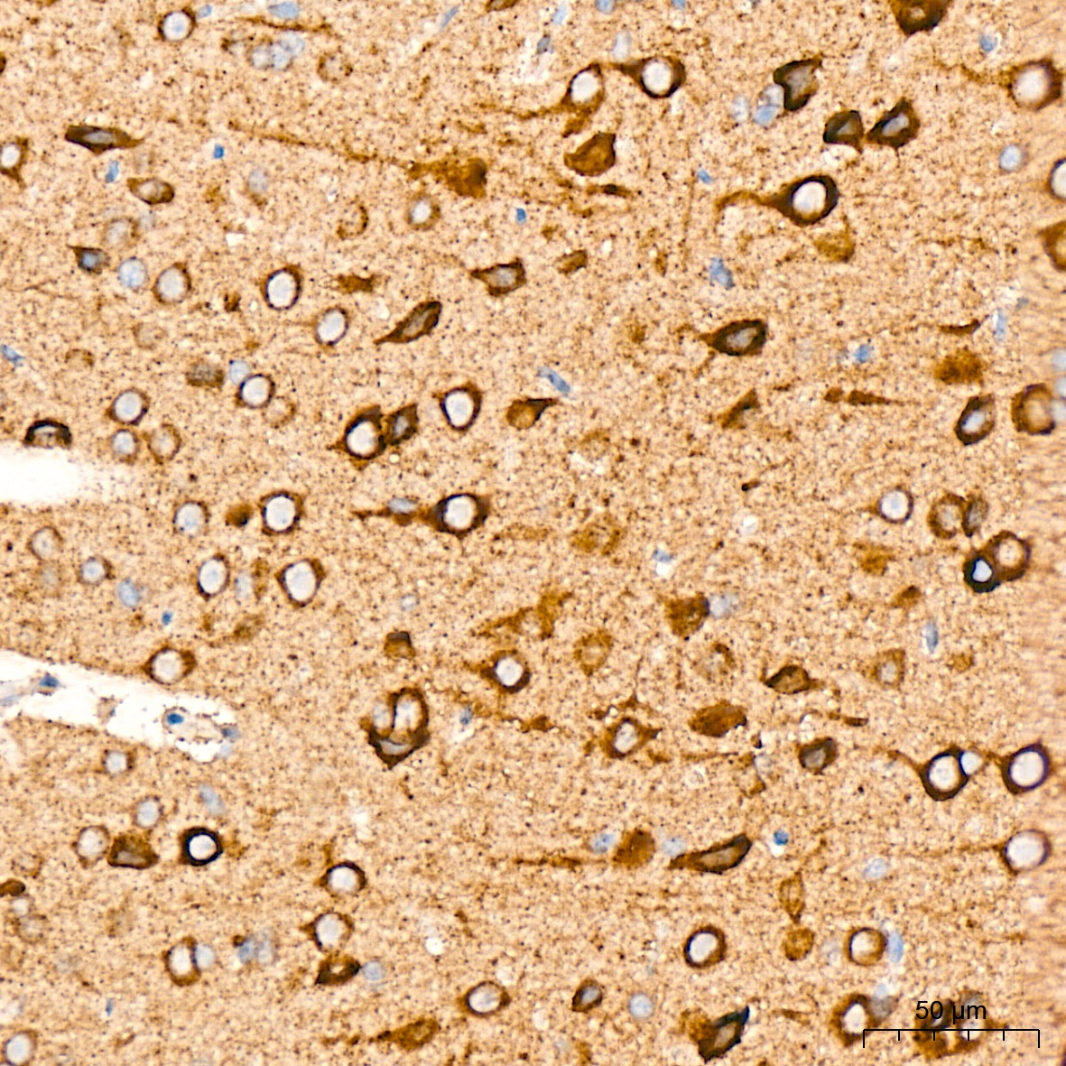
Immunohistochemistry analysis of paraffin-embedded Mouse brain tissue using DCR2 Rabbit mAb (CAB3581) at a dilution of 1:200 (40x lens). High pressure antigen retrieval was performed with 0.01 M citrate buffer (pH 6.0) prior to IHC staining.