The DNA Ligase I Monoclonal Antibody (CAB9301) is a high-quality antibody developed for reliable detection and analysis of target proteins. This antibody, produced in rabbits, exhibits strong reactivity with human samples and is validated for use in Western blot applications. By binding specifically to DNA ligase I, researchers can accurately detect and analyze the enzyme's expression levels in various cell types.DNA ligase I plays a crucial role in maintaining the integrity of the genome by sealing nicks in DNA strands during DNA synthesis and repair.
This antibody is validated for use in WB, IHC-P, IF/ICC, ELISA applications and has demonstrated reactivity against Human, Mouse, Rat samples.
Product Name:
DNA Ligase I Monoclonal Antibody
SKU:
CAB9301
Size:
20μL, 100μL
Reactivity:
Human, Mouse, Rat
Clone Number:
ARC1514
Conjugate:
Unconjugated
Immunogen:
Recombinant protein (or fragment).This information is considered to be commercially sensitive.
Sequence:
Email for sequence
Tested Applications:
WBIHC-PIF/ICCELISA
Recommended Dilution:
WB
1:500 - 1:1000
IHC-P
1:50 - 1:200
IF/ICC
1:50 - 1:200
ELISA
Recommended starting concentration is 1 μg/mL. Please optimize the concentration based on your specific assay requirements.
Synonyms:
LIGI, IMD96, hLig1, DNA Ligase I
Positive Sample:
Jurkat
Cellular Localization:
Cytoplasm, Mitochondrion, Nucleoplasm, Nucleus.
Calculated MW:
102kDa
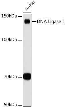
Observed MW:
130kDa
This gene encodes a member of the ATP-dependent DNA ligase protein family. The encoded protein functions in DNA replication, recombination, and the base excision repair process. Mutations in this gene that lead to DNA ligase I deficiency result in immunodeficiency and increased sensitivity to DNA-damaging agents. Disruption of this gene may also be associated with a variety of cancers. Alternative splicing results in multiple transcript variants.
Purification Method
Affinity purification
Gene ID
3978
Buffer Information
Store at -20℃. Avoid freeze / thaw cycles. Buffer: PBS containing 50% glycerol and 0.05% BSA, preserved with proclin300 or sodium azide, pH 7.3.
Western blot analysis of lysates from Jurkat cells, using DNA Ligase I Rabbit mAb (CAB9301) at 1:1000 dilution. Secondary antibody: HRP-conjugated Goat anti-Rabbit IgG (H+L) (CABS014) at 1:10000 dilution. Lysates/proteins: 25μg per lane. Blocking buffer: 3% nonfat dry milk in TBST. Detection: ECL Basic Kit (AbGn00020). Exposure time: 30s.
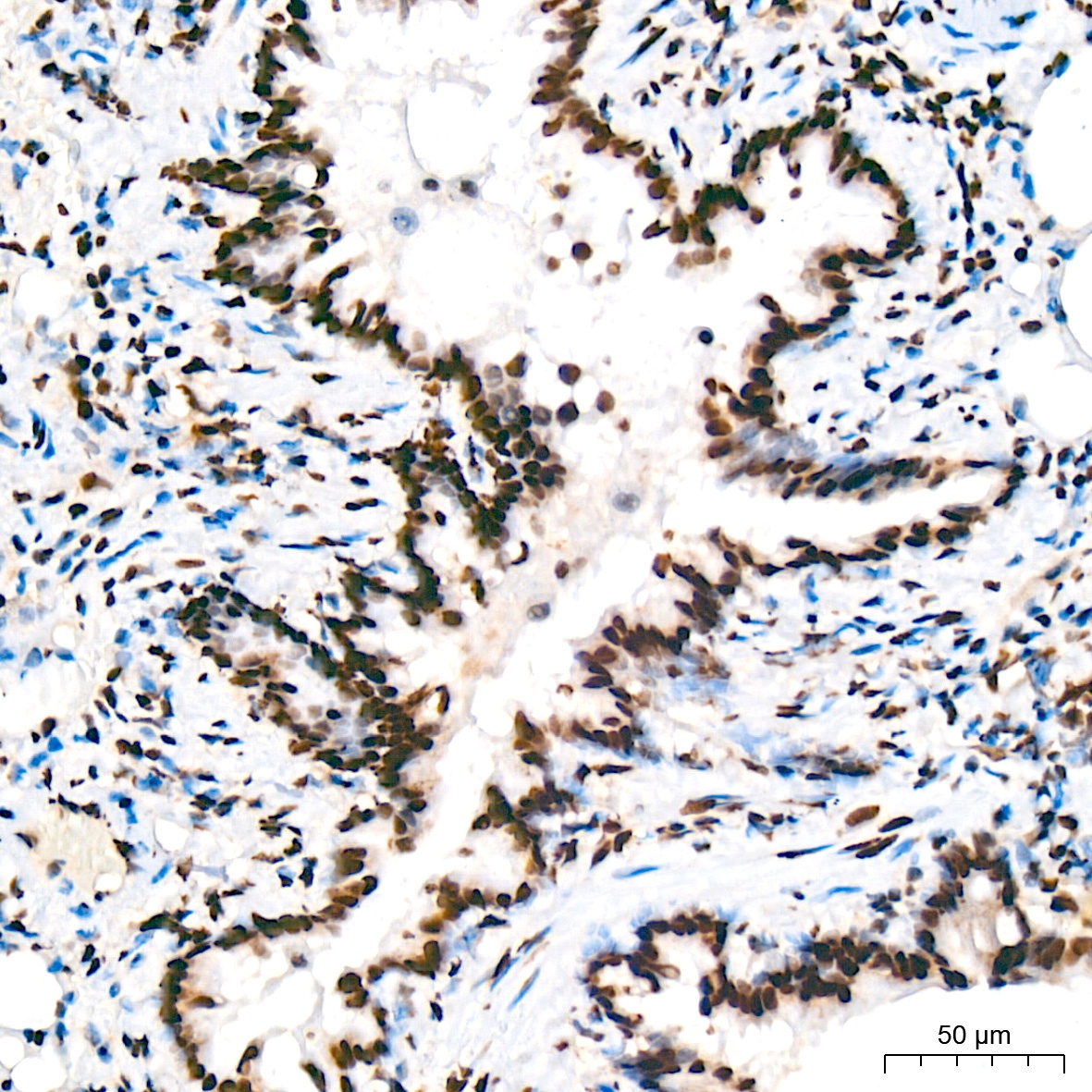
Immunohistochemistry analysis of paraffin-embedded Rat lung tissue using DNA Ligase I Rabbit mAb (CAB9301) at a dilution of 1:200 (40x lens). High pressure antigen retrieval was performed with 0.01 M citrate buffer (pH 6.0) prior to IHC staining.
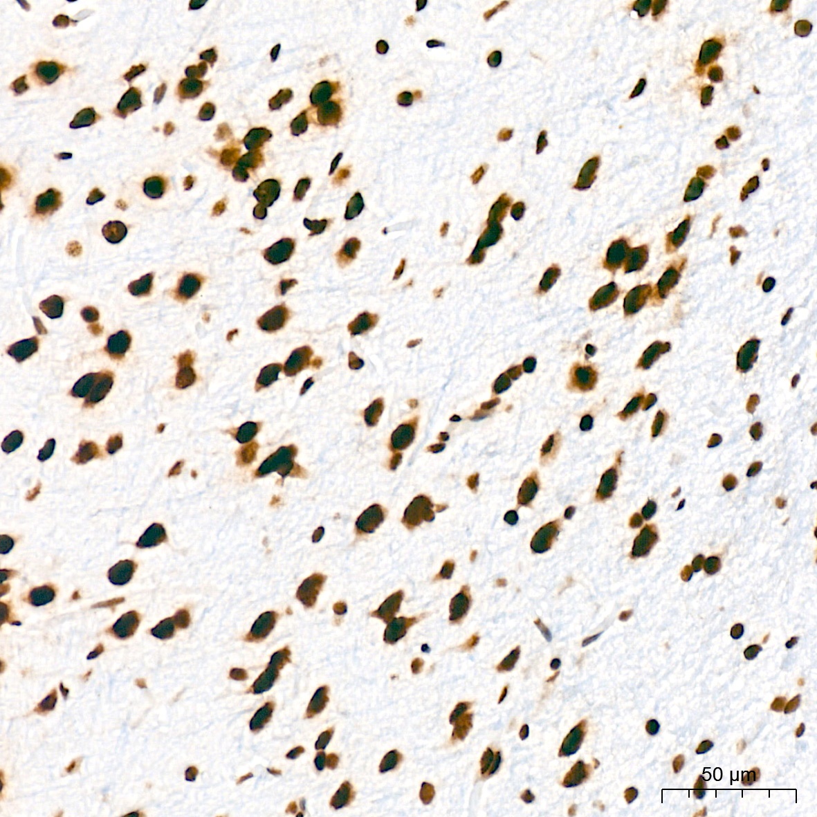
Immunohistochemistry analysis of paraffin-embedded Rat brain tissue using DNA Ligase I Rabbit mAb (CAB9301) at a dilution of 1:200 (40x lens). High pressure antigen retrieval was performed with 0.01 M citrate buffer (pH 6.0) prior to IHC staining.
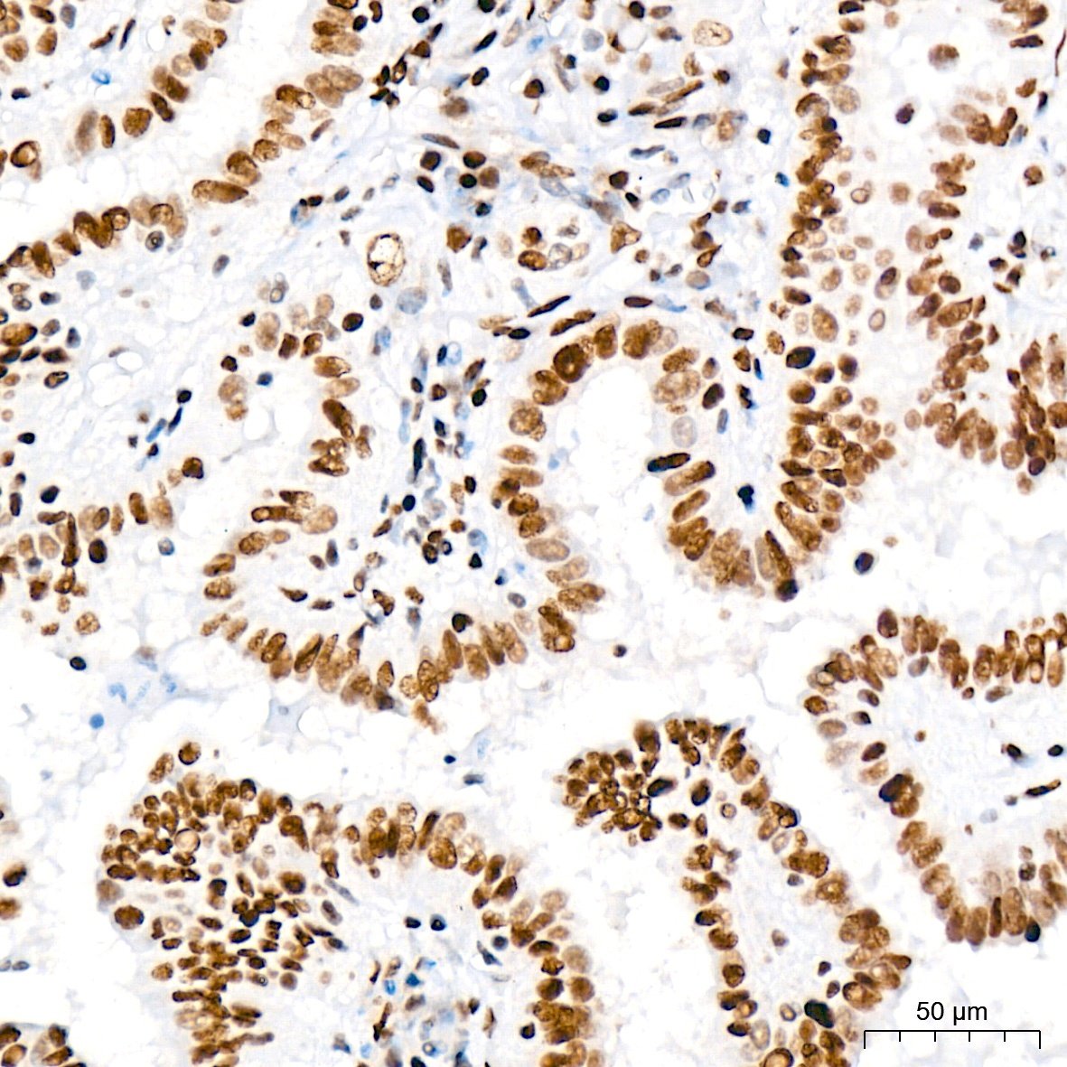
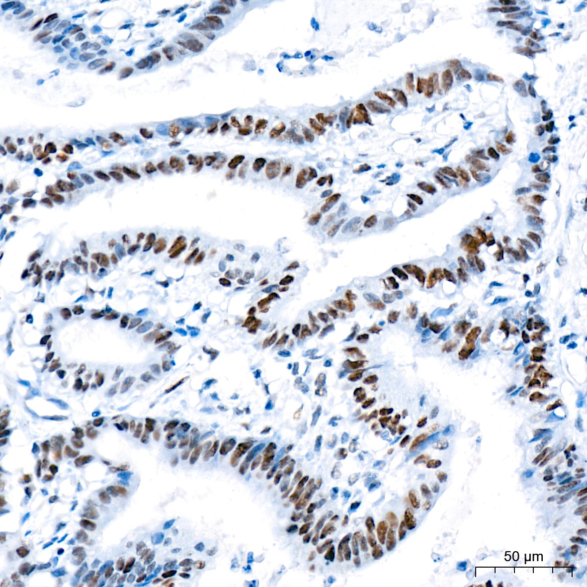
Immunohistochemistry analysis of paraffin-embedded Human thyroid cancer tissue using DNA Ligase I Rabbit mAb (CAB9301) at a dilution of 1:200 (40x lens). High pressure antigen retrieval was performed with 0.01 M citrate buffer (pH 6.0) prior to IHC staining.
Immunohistochemistry analysis of paraffin-embedded Human colon carcinoma tissue using DNA Ligase I Rabbit mAb (CAB9301) at a dilution of 1:200 (40x lens). High pressure antigen retrieval was performed with 0.01 M citrate buffer (pH 6.0) prior to IHC staining.
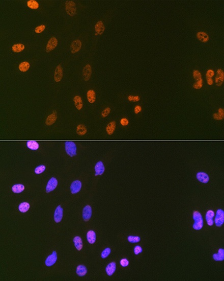
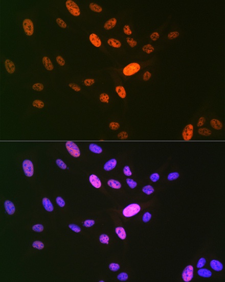
Immunofluorescence analysis of C6 cells using DNA Ligase I Rabbit mAb (CAB9301) at dilution of 1:100 (40x lens). Secondary antibody: Cy3-conjugated Goat anti-Rabbit IgG (H+L) (CABS007) at 1:500 dilution. Blue: DAPI for nuclear staining.
Immunofluorescence analysis of U-2 OS cells using DNA Ligase I Rabbit mAb (CAB9301) at dilution of 1:100 (40x lens). Secondary antibody: Cy3-conjugated Goat anti-Rabbit IgG (H+L) (CABS007) at 1:500 dilution. Blue: DAPI for nuclear staining.


")
 ELISA Kit (HUEB1663)")
 Antibody ELISA Kit (HDES0009)")