The EB3/MAPRE3 Monoclonal Antibody (CAB19768) is a high-quality antibody developed for reliable detection and analysis of target proteins. This antibody, produced in rabbits, is highly specific for human samples and has been validated for use in Western blot applications.EB3, also known as MAPRE3, is essential for regulating microtubule dynamics during cell division and cell migration. Its aberrant expression has been implicated in various diseases, including cancer, neurodegenerative disorders, and developmental abnormalities. The EB3/ MAPRE3 Polyclonal Antibody allows for the detection and analysis of EB3 protein levels in different cell types, making it a valuable tool for researchers in cell biology, cancer research, and neurobiology.
This antibody is validated for use in WB, IF/ICC, ELISA applications and has demonstrated reactivity against Human, Mouse, Rat samples.
Product Name:
EB3/MAPRE3 Monoclonal Antibody
SKU:
CAB19768
Size:
20μL, 100μL
Reactivity:
Human, Mouse, Rat
Clone Number:
ARC2305
Conjugate:
Unconjugated
Immunogen:
Synthetic peptide. This information is considered to be commercially sensitive.
Recommended starting concentration is 1 μg/mL. Please optimize the concentration based on your specific assay requirements.
Synonyms:
EB3, RP3, EBF3, EBF3-S, EB3/MAPRE3
Positive Sample:
Mouse brain, Rat brain
Cellular Localization:
Cytoplasm, Microtubule Cytoskeleton, Microtubule Organizing Center, Perinuclear Region Of Cytoplasm, Spindle Midzone.
Calculated MW:
32kDa
Observed MW:
32kDa
The protein encoded by this gene is a member of the RP/EB family of genes. The protein localizes to the cytoplasmic microtubule network and binds APCL, a homolog of the adenomatous polyposis coli tumor suppressor gene.
Purification Method
Affinity purification
Gene ID
22924
Buffer Information
Store at -20℃. Avoid freeze / thaw cycles. Buffer: PBS containing 50% glycerol and 0.05% BSA, preserved with proclin300 or sodium azide, pH 7.3.
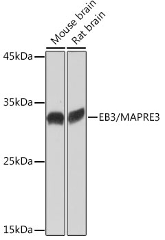
Western blot analysis of various lysates using EB3/MAPRE3 Rabbit mAb (CAB19768) at 1:1000 dilution. Secondary antibody: HRP-conjugated Goat anti-Rabbit IgG (H+L) (CABS014) at 1:10000 dilution. Lysates/proteins: 25μg per lane. Blocking buffer: 3% nonfat dry milk in TBST. Detection: ECL Basic Kit (AbGn00020). Exposure time: 1s.
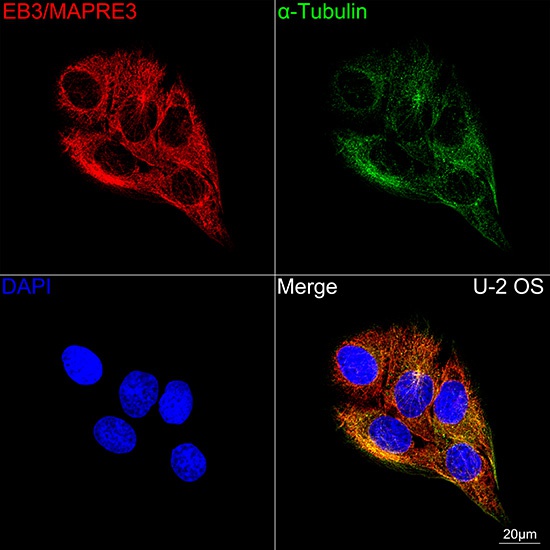
Confocal imaging of U-2 OS cells using EB3/MAPRE3 Rabbit mAb (CAB19768, dilution 1:100) followed by a further incubation with Cy3 Goat Anti-Rabbit IgG (H+L) (CABS007, dilution 1:500) (Red). The cells were counterstained with α-Tubulin Mouse mAb (AC012, dilution 1:400) followed by incubation with ABflo® 488-conjugated Goat Anti-Mouse IgG (H+L) Ab (CABS076, dilution 1:500) (Green). DAPI was used for nuclear staining (Blue). Objective: 100x.
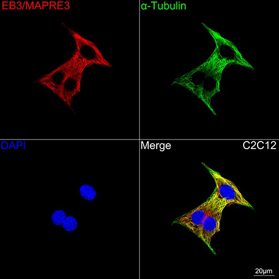
Confocal imaging of C2C12 cells using EB3/MAPRE3 Rabbit mAb (CAB19768, dilution 1:100) followed by a further incubation with Cy3 Goat Anti-Rabbit IgG (H+L) (CABS007, dilution 1:500) (Red). The cells were counterstained with α-Tubulin Mouse mAb (AC012, dilution 1:400) followed by incubation with ABflo® 488-conjugated Goat Anti-Mouse IgG (H+L) Ab (CABS076, dilution 1:500) (Green). DAPI was used for nuclear staining (Blue). Objective: 100x.