The ENT2/SLC29A2 Monoclonal Antibody (CAB19691) is a high-quality antibody developed for reliable detection and analysis of target proteins. The antibody, raised in rabbits, is highly reactive with human samples and has been validated for use in various applications, including Western blot and immunohistochemistry.ENT2 (SLC29A2) plays a crucial role in nucleoside and nucleobase transport across cell membranes, which is essential for cellular processes such as DNA and RNA synthesis. Dysregulation of ENT2 has been linked to various diseases, including cancer and neurodegenerative disorders, making it a promising target for research and drug development.
This antibody is validated for use in IHC-P, IF/ICC, ELISA applications and has demonstrated reactivity against Human, Mouse, Rat samples.
Product Name:
ENT2/SLC29A2 Monoclonal Antibody
SKU:
CAB19691
Size:
20μL, 100μL
Reactivity:
Human, Mouse, Rat
Clone Number:
ARC2234
Conjugate:
Unconjugated
Immunogen:
Synthetic peptide. This information is considered to be commercially sensitive.
The uptake of nucleosides by transporters, such as SLC29A2, is essential for nucleotide synthesis by salvage pathways in cells that lack de novo biosynthetic pathways. Nucleoside transport also plays a key role in the regulation of many physiologic processes through its effect on adenosine concentration at the cell surface (Griffiths et al., 1997 [PubMed 9396714]).
Purification Method
Affinity purification
Gene ID
3177
Buffer Information
Store at -20℃. Avoid freeze / thaw cycles. Buffer: PBS containing 50% glycerol and 0.05% BSA, preserved with proclin300 or sodium azide, pH 7.3.
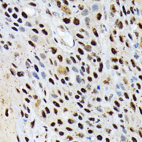
Immunohistochemistry analysis of paraffin-embedded Human esophageal cancer using ENT2/SLC29A2 Rabbit mAb (CAB19691) at dilution of 1:100 (40x lens). Microwave antigen retrieval performed with 0.01M Tris/EDTA Buffer (pH 9.0) prior to IHC staining.
Immunohistochemistry analysis of paraffin-embedded Rat ovary using ENT2/SLC29A2 Rabbit mAb (CAB19691) at dilution of 1:100 (40x lens). Microwave antigen retrieval performed with 0.01M Tris/EDTA Buffer (pH 9.0) prior to IHC staining.
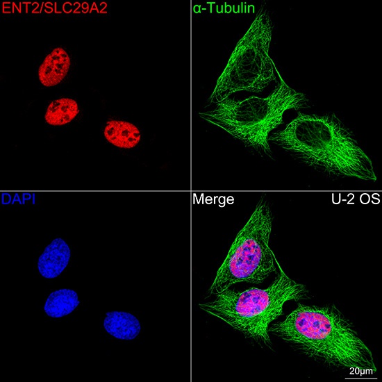
Confocal imaging of U-2 OS cells using ENT2/SLC29A2 Rabbit mAb (CAB19691, dilution 1:200) followed by a further incubation with Cy3 Goat Anti-Rabbit IgG (H+L) (CABS007, dilution 1:500) (Red). The cells were counterstained with α-Tubulin Mouse mAb (AC012, dilution 1:400) followed by incubation with ABflo® 488-conjugated Goat Anti-Mouse IgG (H+L) Ab (CABS076, dilution 1:500) (Green). DAPI was used for nuclear staining (Blue). Objective: 100x.
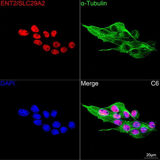
Confocal imaging of C6 cells using ENT2/SLC29A2 Rabbit mAb (CAB19691, dilution 1:200) followed by a further incubation with Cy3 Goat Anti-Rabbit IgG (H+L) (CABS007, dilution 1:500) (Red). The cells were counterstained with α-Tubulin Mouse mAb (AC012, dilution 1:400) followed by incubation with ABflo® 488-conjugated Goat Anti-Mouse IgG (H+L) Ab (CABS076, dilution 1:500) (Green). DAPI was used for nuclear staining (Blue). Objective: 100x.