The Guanylyl Cyclase beta 1 (GUCY1B3) Monoclonal Antibody (CAB3687) is a high-quality antibody developed for reliable detection and analysis of target proteins. This gene encodes the beta subunit of the soluble guanylate cyclase (sGC), which catalyzes the conversion of GTP (guanosine triphosphate) to cGMP (cyclic guanosine monophosphate). The encoded protein contains an HNOX domain, which serves as a receptor for ligands such as nitric oxide, oxygen and nitrovasodilator drugs. Alternative splicing results in multiple transcript variants.
This antibody is validated for use in WB, IHC-P, IF/ICC, ELISA applications and has demonstrated reactivity against Human, Mouse, Rat samples.
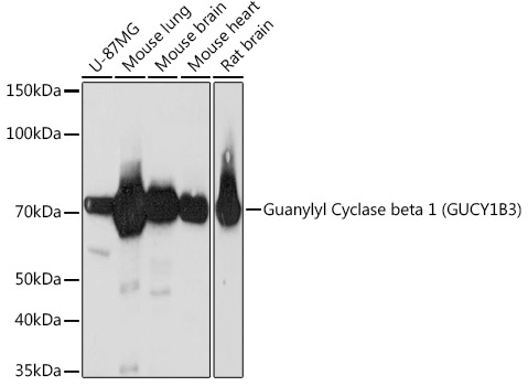
U-87MG, Mouse lung, Mouse brain, Mouse heart, Rat brain
Cellular Localization:
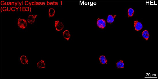
Cytoplasm.
Calculated MW:
71kDa
Observed MW:
71kDa
This gene encodes the beta subunit of the soluble guanylate cyclase (sGC), which catalyzes the conversion of GTP (guanosine triphosphate) to cGMP (cyclic guanosine monophosphate). The encoded protein contains an HNOX domain, which serves as a receptor for ligands such as nitric oxide, oxygen and nitrovasodilator drugs. Alternative splicing results in multiple transcript variants.
Purification Method
Affinity purification
Gene ID
2983
RRID
AB_2863116
Buffer Information
Store at -20℃. Avoid freeze / thaw cycles. Buffer: PBS containing 50% glycerol and 0.05% BSA, preserved with proclin300 or sodium azide, pH 7.3.
Western blot analysis of various lysates using Guanylyl Cyclase beta 1 (GUCY1B3) (GUCY1B3) Rabbit mAb (CAB3687) at 1:1000 dilution. Secondary antibody: HRP-conjugated Goat anti-Rabbit IgG (H+L) (AS014) at 1:10000 dilution. Lysates/proteins: 25μg per lane. Blocking buffer: 3% nonfat dry milk in TBST. Detection: ECL Basic Kit (AbGn00020). Exposure time: 3min.
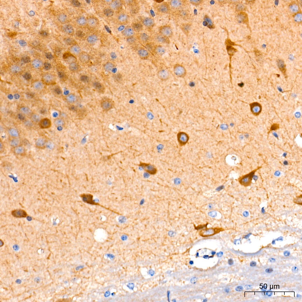
Immunohistochemistry analysis of paraffin-embedded Mouse brain tissue using Guanylyl Cyclase beta 1 (GUCY1B3) Rabbit mAb (CAB3687) at a dilution of 1:200 (40x lens). High pressure antigen retrieval performed with 0.01M Tris-EDTA Buffer (pH 9.0) prior to IHC staining.
Confocal imaging of HEL cells using Guanylyl Cyclase beta 1 (GUCY1B3) Rabbit mAb (CAB3687, dilution 1:200) followed by a further incubation with Cy3 Goat Anti-Rabbit IgG (H+L) (AS007, dilution 1:500) (Red). DAPI was used for nuclear staining (Blue). Objective: 100x.


 ELISA Kit (AEKE01261)")
 ELISA Kit (AEKE02444)")


