
Description
Human IgM Antibody (CAB8677)
The Human IgM Antibody (CAB8677) is a high-quality antibody developed for reliable detection and analysis of target proteins. This polyclonal antibody, generated in rabbits, has been rigorously validated for use in various immunoassays, including Western blot and immunofluorescence applications. It binds specifically to the IgM protein, allowing for precise detection and analysis in a wide range of samples and cell types.
This antibody is validated for use in WB, ELISA applications and has demonstrated reactivity against Human samples.
| Product Name: | Human IgM Antibody |
| SKU: | CAB8677 |
| Size: | 20μL, 100μL |
| Reactivity: | Human |
| Conjugate: | Unconjugated |
| Immunogen: | Recombinant protein (or fragment).This information is considered to be commercially sensitive. | ||||
| Sequence: | MCVP DQDT AIRV FAIP PSFA SIFL TKST KLTC LVTD LTTY DSVT ISWT RQNG EAVK THTN ISES HPNA TFSA VGEA SICE DDWN SGER FTCT VTHT DLPS PLKQ TISR PKGV ALHR PDVY LLPP AREQ LNLR ESAT ITCL VTGF SPAD VFVQ WMQR GQPL SPEK YVTS APMP EPQA PGRY FAHS ILTV SEEE WNTG ETYT CVVA HEAL PNRV TERT VDKS TGKP TLYN VSLV MSDT AGTC Y | ||||
| Tested Applications: | WB ELISA | ||||
| Recommended Dilution: |
| ||||
| Synonyms: | MU, VH, AGM1, Human IgM |
| Positive Sample: | Raji |
| Cellular Localization: | Cell Membrane, Secreted, Single-Pass Type I Membrane Protein. |
| Calculated MW: | 49kDa |
| Observed MW: | 80kDa |
Immunoglobulins (Ig) are the antigen recognition molecules of B cells. An Ig molecule is made up of 2 identical heavy chains and 2 identical light chains (see MIM 147200) joined by disulfide bonds so that each heavy chain is linked to a light chain and the 2 heavy chains are linked together. Each Ig heavy chain has an N-terminal variable (V) region containing the antigen-binding site and a C-terminal constant (C) region, encoded by an individual C region gene, that determines the isotype of the antibody and provides effector or signaling functions. The heavy chain V region is encoded by 1 each of 3 types of genes: V genes (see MIM 147070), joining (J) genes (see MIM 147010), and diversity (D) genes (see MIM 146910). The C region genes are clustered downstream of the V region genes within the heavy chain locus on chromosome 14. The IGHM gene encodes the C region of the mu heavy chain, which defines the IgM isotype. Naive B cells express the transmembrane forms of IgM and IgD (see IGHD; MIM 1471770) on their surface. During an antibody response, activated B cells can switch to the expression of individual downstream heavy chain C region genes by a process of somatic recombination known as isotype switching. In addition, secreted Ig forms that act as antibodies can be produced by alternative RNA processing of the heavy chain C region sequences. Although the membrane forms of all Ig isotypes are monomeric, secreted IgM forms pentamers, and occasionally hexamers, in plasma (summary by Janeway et al., 2005).
| Purification Method | Affinity purification |
| Gene ID | 3507 |
| Buffer Information | Store at -20℃. Avoid freeze / thaw cycles. Buffer: PBS containing 50% glycerol, preserved with proclin300 or sodium azide, pH 7.3. |
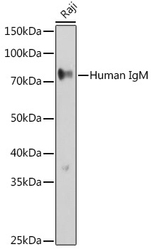 | Western blot analysis of lysates from Raji cells, using Human IgM Rabbit pAb (CAB8677) at 1:500 dilution. Secondary antibody: HRP-conjugated Goat anti-Rabbit IgG (H+L) (CABS014) at 1:10000 dilution. Lysates/proteins: 25μg per lane. Blocking buffer: 3% nonfat dry milk in TBST. Detection: ECL Basic Kit (AbGn00020). Exposure time: 30s. |


 CXXC4 Polyclonal Antibody (PACO44756)")
 CXXC4 Polyclonal Antibody (PACO44756)")
 IL23R Polyclonal Antibody (PACO65154)")
 IL23R Polyclonal Antibody (PACO65154)")
