The iNOS Monoclonal Antibody (CAB3774) is a high-quality antibody developed for reliable detection and analysis of target proteins. This polyclonal antibody, generated in rabbits, shows high reactivity with human iNOS and has been validated for Western blot applications.iNOS is a key player in the production of nitric oxide (NO), a signaling molecule that plays a crucial role in various physiological processes, including host defense, inflammation, and vasodilation. Dysregulation of iNOS and NO production has been implicated in numerous diseases, including cancer, autoimmune disorders, and infectious diseases.
This antibody is validated for use in WB, IF/ICC, ELISA applications and has demonstrated reactivity against Mouse samples.
Product Name:
iNOS Monoclonal Antibody
SKU:
CAB3774
Size:
20μL, 100μL
Reactivity:
Mouse
Clone Number:
ARC0832
Conjugate:
Unconjugated
Immunogen:
Recombinant protein (or fragment).This information is considered to be commercially sensitive.
Recommended starting concentration is 1 μg/mL. Please optimize the concentration based on your specific assay requirements.
Synonyms:
NOS, INOS, NOS2A, HEP-NOS, iNOS
Positive Sample:
RAW 264.7 treated with LPS, NR8383 treated with LPS
Cellular Localization:
Cortical Cytoskeleton, Cytoplasm, Cytosol, Nucleoplasm, Nucleus, Perinuclear Region Of Cytoplasm, Peroxisomal Matrix, Peroxisome, Plasma Membrane.
Calculated MW:
131kDa
Observed MW:
131kDa
Nitric oxide is a reactive free radical which acts as a biologic mediator in several processes, including neurotransmission and antimicrobial and antitumoral activities. This gene encodes a nitric oxide synthase which is expressed in liver and is inducible by a combination of lipopolysaccharide and certain cytokines. Three related pseudogenes are located within the Smith-Magenis syndrome region on chromosome 17.
Purification Method
Affinity purification
Gene ID
4843
RRID
AB_3094627
Buffer Information
Store at -20℃. Avoid freeze / thaw cycles. Buffer: PBS containing 50% glycerol and 0.05% BSA, preserved with proclin300 or sodium azide, pH 7.3.
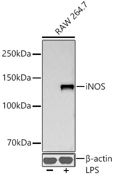
Western blot analysis of lysates from RAW 264.7 cells using iNOS Rabbit mAb (CAB3774) at 1:1000 dilution incubated overnight at 4℃. Raw264.7 cells were treated with LPS (1 μg/ml) at 37℃ for 8 hours. Secondary antibody: HRP-conjugated Goat anti-Rabbit IgG (H+L) (CABS014) at 1:10000 dilution. Lysates/proteins: 30 μg per lane. Blocking buffer: 3% nonfat dry milk in TBST. Detection: ECL Basic Kit (AbGn00020). Exposure time: 30 s.
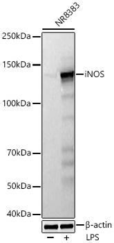
Western blot analysis of lysates from NR8383 cells using iNOS Rabbit mAb (CAB3774) at 1:1000 dilution incubated at room temperature for 1.5 hours. NR8383 cells were treated with LPS (1 ug/mL) for 8 hours. Secondary antibody: HRP-conjugated Goat anti-Rabbit IgG (H+L) (CABS014) at 1:10000 dilution. Lysates/proteins: 30 μg per lane. Blocking buffer: 3% nonfat dry milk in TBST. Detection: ECL Basic Kit (AbGn00020). Exposure time: 5 s.