The iNOS Monoclonal Antibody (CAB3774) is a high-quality antibody developed for reliable detection and analysis of target proteins. Nitric oxide is a reactive free radical which acts as a biologic mediator in several processes, including neurotransmission and antimicrobial and antitumoral activities. This gene encodes a nitric oxide synthase which is expressed in liver and is inducible by a combination of lipopolysaccharide and certain cytokines. Three related pseudogenes are located within the Smith-Magenis syndrome region on chromosome 17.
This antibody is validated for use in WB, IF/ICC, ELISA applications and has demonstrated reactivity against Mouse samples.
Product Name:
iNOS Monoclonal Antibody
SKU:
CAB3774
Size:
100μL, 20μL
Reactivity:
Mouse
Clone Number:
ARC0832
Conjugate:
Unconjugated
Immunogen:
Recombinant protein (or fragment).This information is considered to be commercially sensitive.
Tested Applications:
WBIF/ICCELISA
Recommended Dilution:
WB
1:1000 - 1:4000
IF/ICC
1:50 - 1:200
ELISA
Recommended starting concentration is 1 μg/mL. Please optimize the concentration based on your specific assay requirements.
Synonyms:
NOS, INOS, NOS2A, HEP-NOS, iNOS
Positive Sample:
RAW 264.7 treated with LPS, NR8383 treated with LPS
Cellular Localization:
Cortical Cytoskeleton, Cytoplasm, Cytosol, Nucleoplasm, Nucleus, Perinuclear Region Of Cytoplasm, Peroxisomal Matrix, Peroxisome, Plasma Membrane.
Calculated MW:
131 kDa
Observed MW:
131 kDa
Nitric oxide is a reactive free radical which acts as a biologic mediator in several processes, including neurotransmission and antimicrobial and antitumoral activities. This gene encodes a nitric oxide synthase which is expressed in liver and is inducible by a combination of lipopolysaccharide and certain cytokines. Three related pseudogenes are located within the Smith-Magenis syndrome region on chromosome 17.
Purification Method
Affinity purification
Gene ID
4843
RRID
AB_3094627
Buffer Information
Store at -20℃. Avoid freeze / thaw cycles. Buffer: PBS containing 50% glycerol and 0.05% BSA, preserved with proclin300 or sodium azide, pH 7.3.
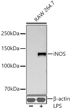
Western blot analysis of lysates from RAW 264.7 cells using iNOS Rabbit mAb (CAB3774) at 1:1000 dilution incubated overnight at 4℃. Raw264.7 cells were treated with LPS (1 μg/ml) at 37℃ for 8 hours. Secondary antibody: HRP-conjugated Goat anti-Rabbit IgG (H+L) (AS014) at 1:10000 dilution. Lysates/proteins: 30 μg per lane. Blocking buffer: 3% nonfat dry milk in TBST. Detection: ECL Basic Kit (AbGn00020). Exposure time: 30 s.
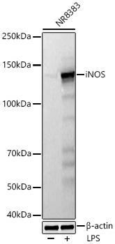
Western blot analysis of lysates from NR8383 cells using iNOS Rabbit mAb (CAB3774) at 1:1000 dilution incubated at room temperature for 1.5 hours. NR8383 cells were treated with LPS (1 ug/mL) for 8 hours. Secondary antibody: HRP-conjugated Goat anti-Rabbit IgG (H+L) (AS014) at 1:10000 dilution. Lysates/proteins: 30 μg per lane. Blocking buffer: 3% nonfat dry milk in TBST. Detection: ECL Basic Kit (AbGn00020). Exposure time: 5 s.