The KAT8/MYST1/MOF Monoclonal Antibody (CAB3390) is a high-quality antibody developed for reliable detection and analysis of target proteins. This antibody, produced in rabbits, is highly specific to human samples and has been validated for use in Western blot applications. By binding to the KAT8/MYST1/MOF protein, this antibody enables accurate detection and analysis in various cell types, making it ideal for research in epigenetics and cancer biology.KAT8/MYST1/MOF is a histone acetyltransferase that catalyzes the acetylation of histone H4, leading to changes in chromatin structure and gene expression.
This antibody is validated for use in IHC-P, IF/ICC, ELISA, IF-P applications and has demonstrated reactivity against Human, Rat samples.
Product Name:
KAT8/MYST1/MOF Monoclonal Antibody
SKU:
CAB3390
Size:
20μL, 100μL
Reactivity:
Human, Rat
Clone Number:
ARC1964
Conjugate:
Unconjugated
Immunogen:
Recombinant protein (or fragment).This information is considered to be commercially sensitive.
Recommended starting concentration is 1 μg/mL. Please optimize the concentration based on your specific assay requirements.
Synonyms:
MOF, hMOF, MYST1, LIGOWS, ZC2HC8, KAT8/MYST1/MOF
Cellular Localization:
Chromosome, Nucleus.
Calculated MW:
52kDa
Observed MW:
Refertofigures
This gene encodes a member of the MYST histone acetylase protein family. The encoded protein has a characteristic MYST domain containing an acetyl-CoA-binding site, a chromodomain typical of proteins which bind histones, and a C2HC-type zinc finger. Multiple transcript variants encoding different isoforms have been found for this gene.
Purification Method
Affinity purification
Gene ID
84148
RRID
AB_2863049
Buffer Information
Store at -20℃. Avoid freeze / thaw cycles. Buffer: PBS containing 50% glycerol and 0.05% BSA, preserved with proclin300 or sodium azide, pH 7.3.
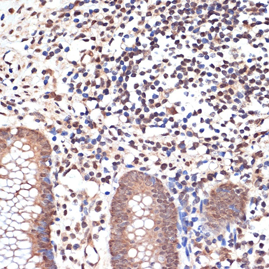
Immunohistochemistry analysis of paraffin-embedded Human appendix using KAT8/MYST1/MOF Rabbit mAb (CAB3390) at dilution of 1:100 (40x lens). Microwave antigen retrieval performed with 0.01M Tris/EDTA Buffer (pH 9.0) prior to IHC staining.
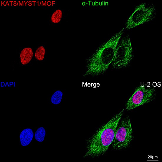
Confocal imaging of U-2 OS cells using KAT8/MYST1/MOF Rabbit mAb (CAB3390,dilution 1:200) followed by a further incubation with Cy3 Goat Anti-Rabbit IgG (H+L) (CABS007,dilution 1:500)(Red).The cells were counterstained with α-Tubulin Mouse mAb (AC012, dilution 1:400) followed by incubation with ABflo® 488-conjugated Goat Anti-Mouse IgG (H+L) Ab (CABS076, dilution 1:500) (Green).DAPI was used for nuclear staining (Blue). Objective: 100x.


")
")


