The KAT8/MYST1/MOF Monoclonal Antibody (CAB3390) is a high-quality antibody developed for reliable detection and analysis of target proteins. This gene encodes a member of the MYST histone acetylase protein family. The encoded protein has a characteristic MYST domain containing an acetyl-CoA-binding site, a chromodomain typical of proteins which bind histones, and a C2HC-type zinc finger. Multiple transcript variants encoding different isoforms have been found for this gene.
This antibody is validated for use in IHC-P, IF/ICC, ELISA, IF-P applications and has demonstrated reactivity against Human, Rat samples.
Product Name:
KAT8/MYST1/MOF Monoclonal Antibody
SKU:
CAB3390
Size:
100μL, 20μL
Reactivity:
Human, Rat
Clone Number:
ARC1964
Conjugate:
Unconjugated
Immunogen:
Recombinant protein (or fragment).This information is considered to be commercially sensitive.
Tested Applications:
IHC-PIF/ICCELISAIF-P
Recommended Dilution:
IF/ICC
1:200 - 1:800
IF-P
1:200 - 1:800
IHC-P
1:100 - 1:1000
ELISA
Recommended starting concentration is 1 μg/mL. Please optimize the concentration based on your specific assay requirements.
Synonyms:
MOF, hMOF, MYST1, LIGOWS, ZC2HC8, KAT8/MYST1/MOF
Cellular Localization:
Chromosome, Nucleus.
Calculated MW:
52kDa
Observed MW:
Refer to figures
This gene encodes a member of the MYST histone acetylase protein family. The encoded protein has a characteristic MYST domain containing an acetyl-CoA-binding site, a chromodomain typical of proteins which bind histones, and a C2HC-type zinc finger. Multiple transcript variants encoding different isoforms have been found for this gene.
Purification Method
Affinity purification
Gene ID
84148
RRID
AB_2863049
Buffer Information
Store at -20℃. Avoid freeze / thaw cycles. Buffer: PBS containing 50% glycerol and 0.05% BSA, preserved with proclin300 or sodium azide, pH 7.3.
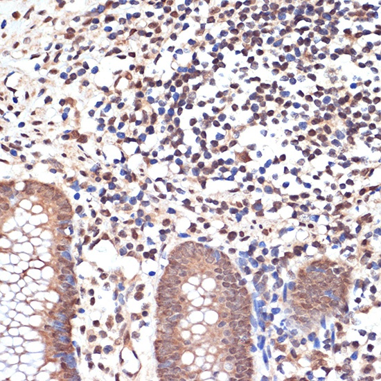
Immunohistochemistry analysis of paraffin-embedded Human appendix using KAT8/MYST1/MOF Rabbit mAb (CAB3390) at dilution of 1:100 (40x lens). Microwave antigen retrieval performed with 0.01M Tris/EDTA Buffer (pH 9.0) prior to IHC staining.
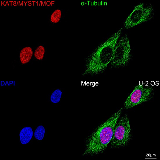
Confocal imaging of U-2 OS cells using KAT8/MYST1/MOF Rabbit mAb (CAB3390,dilution 1:200) followed by a further incubation with Cy3 Goat Anti-Rabbit IgG (H+L) (AS007,dilution 1:500)(Red).The cells were counterstained with α-Tubulin Mouse mAb (AC012, dilution 1:400) followed by incubation with ABflo® 488-conjugated Goat Anti-Mouse IgG (H+L) Ab (AS076, dilution 1:500) (Green).DAPI was used for nuclear staining (Blue). Objective: 100x.


")


