The LLGL1 Antibody (CAB20205) is a high-quality antibody developed for reliable detection and analysis of target proteins. This antibody, raised in rabbits, exhibits high reactivity with human samples and is validated for use in Western blot applications.LLGL1, or lethal giant larvae homolog 1, is a key regulator of cell polarity and asymmetric cell division, important for maintaining tissue organization and preventing tumor formation. By targeting the LLGL1 protein, researchers can gain insights into its function in various cell types and its implications in cancer biology.
This antibody is validated for use in WB, IHC-P, IF/ICC, ELISA applications and has demonstrated reactivity against Human, Mouse, Rat samples.
Product Name:
LLGL1 Antibody
SKU:
CAB20205
Size:
20μL, 100μL
Reactivity:
Human, Mouse, Rat
Conjugate:
Unconjugated
Immunogen:
Recombinant protein (or fragment).This information is considered to be commercially sensitive.
This gene encodes a protein that is similar to a tumor suppressor in Drosophila. The protein is part of a cytoskeletal network and is associated with nonmuscle myosin II heavy chain and a kinase that specifically phosphorylates this protein at serine residues. The gene is located within the Smith-Magenis syndrome region on chromosome 17.
Purification Method
Affinity purification
Gene ID
3996
Buffer Information
Store at -20℃. Avoid freeze / thaw cycles. Buffer: PBS containing 50% glycerol, preserved with proclin300 or sodium azide, pH 7.3.
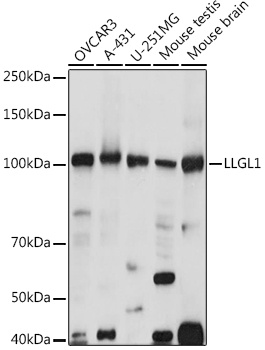
Western blot analysis of various lysates using LLGL1 Rabbit pAb (CAB20205) at 1:1000 dilution. Secondary antibody: HRP-conjugated Goat anti-Rabbit IgG (H+L) (CABS014) at 1:10000 dilution. Lysates/proteins: 25μg per lane. Blocking buffer: 3% nonfat dry milk in TBST. Detection: ECL Basic Kit (AbGn00020). Exposure time: 10s.
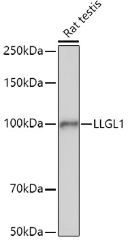
Western blot analysis of lysates from Rat testis, using LLGL1 Rabbit pAb (CAB20205) at 1:1000 dilution. Secondary antibody: HRP-conjugated Goat anti-Rabbit IgG (H+L) (CABS014) at 1:10000 dilution. Lysates/proteins: 25μg per lane. Blocking buffer: 3% nonfat dry milk in TBST. Detection: ECL Basic Kit (AbGn00020). Exposure time: 60s.
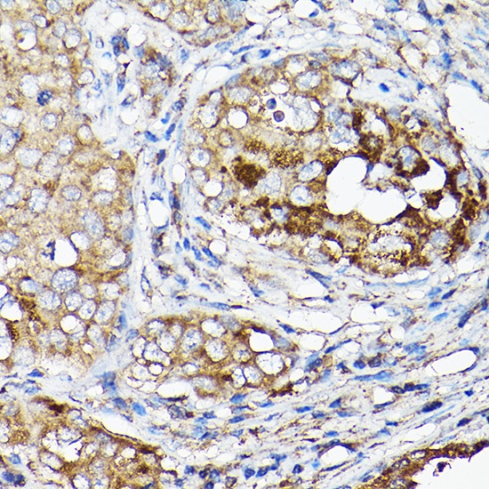
Immunohistochemistry analysis of paraffin-embedded Human breast cancer using LLGL1 Rabbit pAb (CAB20205) at dilution of 1:50 (40x lens). High pressure antigen retrieval performed with 0.01M Citrate buffer (pH 6.0) prior to IHC staining.
Immunofluorescence analysis of A-549 cells using LLGL1 Rabbit pAb (CAB20205) at dilution of 1:100. Secondary antibody: Cy3-conjugated Goat anti-Rabbit IgG (H+L) (CABS007) at 1:500 dilution. Blue: DAPI for nuclear staining.