The MDA5 Monoclonal Antibody (CAB2419) is a high-quality antibody developed for reliable detection and analysis of target proteins. This antibody is produced in rabbits and is highly reactive with human samples, making it ideal for use in various research applications such as Western blotting.MDA5 is a cytoplasmic pattern recognition receptor involved in the detection of viral RNA, playing a crucial role in antiviral immune responses. Dysregulation of MDA5 has been associated with autoimmune diseases such as dermatomyositis and interstitial lung disease.
This antibody is validated for use in WB, ELISA applications and has demonstrated reactivity against Human, Mouse, Rat samples.
Product Name:
MDA5 Monoclonal Antibody
SKU:
CAB2419
Size:
20μL, 100μL
Reactivity:
Human, Mouse, Rat
Clone Number:
ARC0760
Conjugate:
Unconjugated
Immunogen:
Synthetic peptide. This information is considered to be commercially sensitive.
IFIH1 encodes MDA5 which is an intracellular sensor of viral RNA that triggers the innate immune response. Sensing RNA length and secondary structure, MDA5 binds dsRNA oligonucleotides with a modified DExD/H-box helicase core and a C-terminal domain, thus leading to a proinflammatory response that includes interferons. It has been shown that Coronaviruses (CoVs) as well as various other virus families, are capable of evading the MDA5-dependent interferon response, thus impeding the activation of the innate immune response to infection. MDA5 has also been shown to play an important role in enhancing natural killer cell function in malaria infection. In addition to its protective role in antiviral responses, MDA5 has been implicated in autoimmune and autoinflammatory diseases such as type 1 diabetes, systemic lupus erythematosus, and Aicardi-Goutieres syndrome
Purification Method
Affinity purification
Gene ID
64135
Buffer Information
Store at -20℃. Avoid freeze / thaw cycles. Buffer: PBS containing 50% glycerol and 0.05% BSA, preserved with proclin300 or sodium azide, pH 7.3.
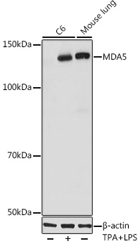
Western blot analysis of various lysates using MDA5 Rabbit mAb (CAB2419) at 1:1000 dilution. C6 cells were treated with LPS for 6 hours and TPA for 3 hours of stimulation. Secondary antibody: HRP-conjugated Goat anti-Rabbit IgG (H+L) (CABS014) at 1:10000 dilution. Lysates/proteins: 25μg per lane. Blocking buffer: 3% nonfat dry milk in TBST. Detection: ECL Basic Kit (AbGn00020). Exposure time: 10s.
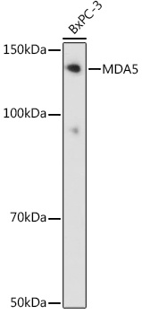
Western blot analysis of lysates from BxPC-3 cells, using MDA5 Rabbit mAb (CAB2419) at 1:1000 dilution. Secondary antibody: HRP-conjugated Goat anti-Rabbit IgG (H+L) (CABS014) at 1:10000 dilution. Lysates/proteins: 25μg per lane. Blocking buffer: 3% nonfat dry milk in TBST. Detection: ECL Basic Kit (AbGn00020). Exposure time: 90s.