The hnRNP A1 Monoclonal Antibody (CAB11564) is a high-quality antibody developed for reliable detection and analysis of target proteins. This high-quality antibody, produced in rabbits, is specifically designed for use in Western blot applications and is highly reactive with human samples.HNRNP A1, also known as heterogeneous nuclear ribonucleoprotein A1, is involved in various cellular processes, including pre-mRNA splicing, mRNA stability, and translation. Dysregulation of HNRNP A1 has been linked to several diseases, including cancer and neurodegenerative disorders, making it an important target for research in these areas.
This antibody is validated for use in WB, IHC-P, IF/ICC, IP, ELISA applications and has demonstrated reactivity against Human, Mouse, Rat samples.
Product Name:
hnRNP A1 Monoclonal Antibody
SKU:
CAB11564
Size:
20μL, 100μL
Reactivity:
Human, Mouse, Rat
Clone Number:
ARC0633
Conjugate:
Unconjugated
Immunogen:
Synthetic peptide. This information is considered to be commercially sensitive.
0.5μg-4μg antibody for 200μg-400μg extracts of whole cells
ELISA
Recommended starting concentration is 1 μg/mL. Please optimize the concentration based on your specific assay requirements.
Synonyms:
UP 1, ALS19, ALS20, HNRPA1, IBMPFD3, HNRPA1L3, hnRNP A1, hnRNP-A1
Positive Sample:
HeLa, 293T, HepG2, Mouse brain, Rat brain
Cellular Localization:
Cytoplasm, Nucleus.
Calculated MW:
39kDa
Observed MW:
30kDa/34kDa/39kDa
This gene encodes a member of a family of ubiquitously expressed heterogeneous nuclear ribonucleoproteins (hnRNPs), which are RNA-binding proteins that associate with pre-mRNAs in the nucleus and influence pre-mRNA processing, as well as other aspects of mRNA metabolism and transport. The protein encoded by this gene is one of the most abundant core proteins of hnRNP complexes and plays a key role in the regulation of alternative splicing. Mutations in this gene have been observed in individuals with amyotrophic lateral sclerosis 20. Multiple alternatively spliced transcript variants have been found. There are numerous pseudogenes of this gene distributed throughout the genome.hnRNP A1 has three isoforms with MW 30 kDa, 34 kDa and 39 kDa.
Purification Method
Affinity purification
Gene ID
3178
RRID
AB_2861599
Buffer Information
Store at -20℃. Avoid freeze / thaw cycles. Buffer: PBS containing 50% glycerol and 0.05% BSA, preserved with proclin300 or sodium azide, pH 7.3.
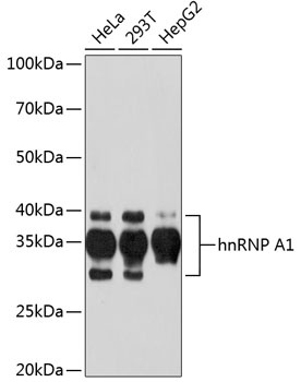
Western blot analysis of various lysates using hnRNP A1 Rabbit mAb (CAB11564) at 1:1000 dilution. Secondary antibody: HRP-conjugated Goat anti-Rabbit IgG (H+L) (CABS014) at 1:10000 dilution. Lysates/proteins: 25μg per lane. Blocking buffer: 3% nonfat dry milk in TBST. Detection: ECL Basic Kit (AbGn00020). Exposure time: 60s.
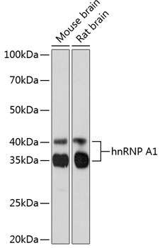
Western blot analysis of various lysates using hnRNP A1 Rabbit mAb (CAB11564) at 1:1000 dilution. Secondary antibody: HRP-conjugated Goat anti-Rabbit IgG (H+L) (CABS014) at 1:10000 dilution. Lysates/proteins: 25μg per lane. Blocking buffer: 3% nonfat dry milk in TBST. Detection: ECL Basic Kit (AbGn00020). Exposure time: 3min.
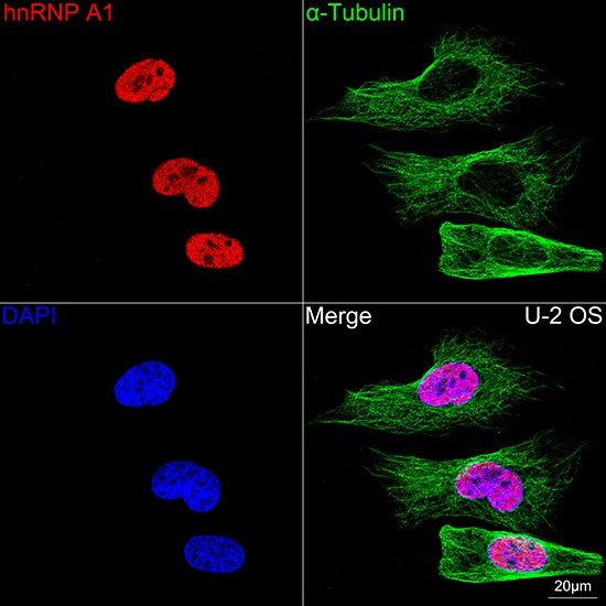
Confocal imaging of U-2 OS cells using hnRNP A1 Rabbit mAb (CAB11564,dilution 1:200) followed by a further incubation with Cy3 Goat Anti-Rabbit IgG (H+L) (CABS007,dilution 1:500)(Red).The cells were counterstained with α-Tubulin Mouse mAb (AC012, dilution 1:400) followed by incubation with ABflo® 488-conjugated Goat Anti-Mouse IgG (H+L) Ab (CABS076, dilution 1:500) (Green).DAPI was used for nuclear staining (Blue). Objective: 100x.
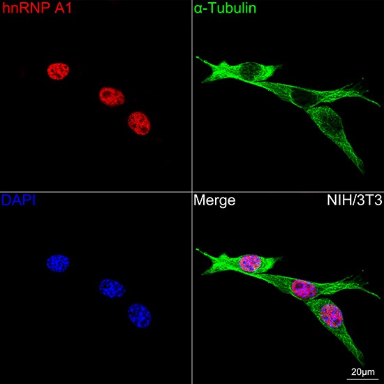
Confocal imaging of NIH/3T3 cells using hnRNP A1 Rabbit mAb (CAB11564,dilution 1:200) followed by a further incubation with Cy3 Goat Anti-Rabbit IgG (H+L) (CABS007,dilution 1:500)(Red).The cells were counterstained with α-Tubulin Mouse mAb (AC012, dilution 1:400) followed by incubation with ABflo® 488-conjugated Goat Anti-Mouse IgG (H+L) Ab (CABS076, dilution 1:500) (Green).DAPI was used for nuclear staining (Blue). Objective: 100x.
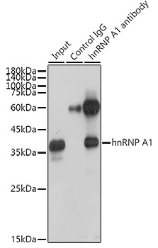
Immunoprecipitation analysis of 300 μg extracts of HeLa cells using 3 μg hnRNP A1 antibody (CAB11564). Western blot was performed from the immunoprecipitate using hnRNP A1 antibody (CAB11564) at a dilution of 1:1000.