The MPC2 Antibody (CAB20196) is a high-quality antibody developed for reliable detection and analysis of target proteins. This antibody, produced in rabbits, is highly specific to human samples and has been validated for use in Western blot applications. By targeting the MPC2 protein, this antibody enables the accurate detection and analysis of this critical mitochondrial transporter in various cell types.MPC2 is a key player in cellular metabolism, specifically in the transport of pyruvate into the mitochondria for energy production.
This antibody is validated for use in WB, IHC-P, ELISA applications and has demonstrated reactivity against Mouse, Rat samples.
Product Name:
MPC2 Antibody
SKU:
CAB20196
Size:
20μL, 100μL
Reactivity:
Mouse, Rat
Conjugate:
Unconjugated
Immunogen:
Synthetic peptide. This information is considered to be commercially sensitive.
Recommended starting concentration is 1 μg/mL. Please optimize the concentration based on your specific assay requirements.
Synonyms:
BRP44, SLC54A2, MPC2
Positive Sample:
Mouse kidney
Cellular Localization:
Inner Mitochondrial Membrane Protein Complex, Mitochondrial Inner Membrane, Mitochondrion, Nucleus.
Calculated MW:
14kDa
Observed MW:
14kDa
Enables identical protein binding activity. Predicted to be involved in mitochondrial pyruvate transmembrane transport. Predicted to act upstream of or within mitochondrial acetyl-CoA biosynthetic process from pyruvate and positive regulation of insulin secretion involved in cellular response to glucose stimulus. Located in mitochondrion.
Purification Method
Affinity purification
Gene ID
25874
Buffer Information
Store at -20℃. Avoid freeze / thaw cycles. Buffer: PBS containing 50% glycerol, preserved with proclin300 or sodium azide, pH 7.3.
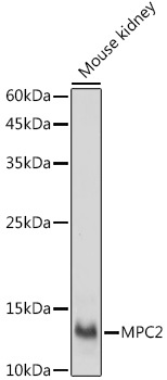
Western blot analysis of lysates from Mouse kidney, using MPC2 Rabbit pAb (CAB20196) at 1:1000 dilution. Secondary antibody: HRP-conjugated Goat anti-Rabbit IgG (H+L) (CABS014) at 1:10000 dilution. Lysates/proteins: 25μg per lane. Blocking buffer: 3% nonfat dry milk in TBST. Detection: ECL Basic Kit (AbGn00020). Exposure time: 30s.
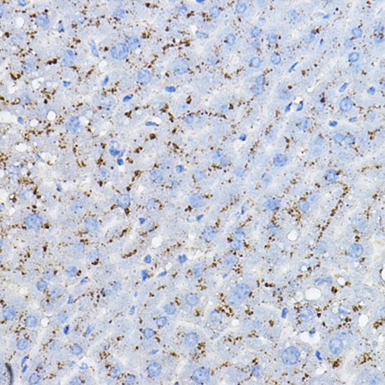
Immunohistochemistry analysis of paraffin-embedded Mouse liver using MPC2 Rabbit pAb (CAB20196) at dilution of 1:100 (40x lens). High pressure antigen retrieval performed with 0.01M Citrate buffer (pH 6.0) prior to IHC staining.